Confirming a diagnosis of liver cirrhosis involves a multi-stage clinical process designed to assess the extent of liver scarring and the organ’s remaining functional capacity. Because cirrhosis represents the advanced stage of various chronic liver conditions, clinicians in the United Kingdom use a combination of laboratory blood tests, sophisticated imaging technologies, and sometimes tissue sampling to reach a definitive conclusion. These investigations are essential not only for identifying the presence of permanent scar tissue but also for determining the underlying cause and monitoring for potential complications such as portal hypertension or liver cancer. Early and accurate confirmation allows healthcare teams to implement management plans aimed at stabilising the liver and preventing further deterioration. While many tests can suggest the presence of liver damage, a formal confirmation usually requires evidence of structural changes that have permanently altered the liver’s architecture.
What We’ll Discuss in This Article
- The role of liver function and specialised blood tests in assessing damage.
- How ultrasound and CT scans identify structural changes in the liver.
- The importance of transient elastography in measuring liver stiffness.
- When a liver biopsy is necessary to confirm the diagnosis.
- Endoscopic investigations for complications related to cirrhosis.
- The tiered approach used by the NHS to reach a definitive diagnosis.
Initial Laboratory Investigations and Blood Markers
Initial blood tests provide the first clinical clues of liver cirrhosis by measuring specific enzymes, proteins, and clotting factors that reflect the organ’s overall health. While no single blood test can definitively confirm cirrhosis on its own, certain patterns in the results are highly suggestive of advanced scarring. The NHS states that a GP may suspect cirrhosis based on your symptoms and a blood test called a liver function test to check how well your liver is working.
In a cirrhotic liver, the production of essential proteins like albumin often decreases, while the time it takes for blood to clot, measured by the Prothrombin Time or International Normalised Ratio, may increase. Clinicians also look at the platelet count; a low platelet count is a common indicator that scarring has begun to affect blood flow through the liver. These markers, when combined with elevated liver enzymes, help create a biochemical profile that guides the next steps in the diagnostic pathway.
Non-Invasive Imaging and Structural Assessment
Imaging scans are a fundamental part of the diagnostic process as they allow doctors to visualise the physical changes to the liver’s shape and texture. Ultrasound is typically the first imaging tool used, as it can detect if the liver has become small, firm, or lumpy, which are classic signs of cirrhosis. It also allows for the assessment of blood flow in the portal vein and the detection of fluid buildup in the abdomen.
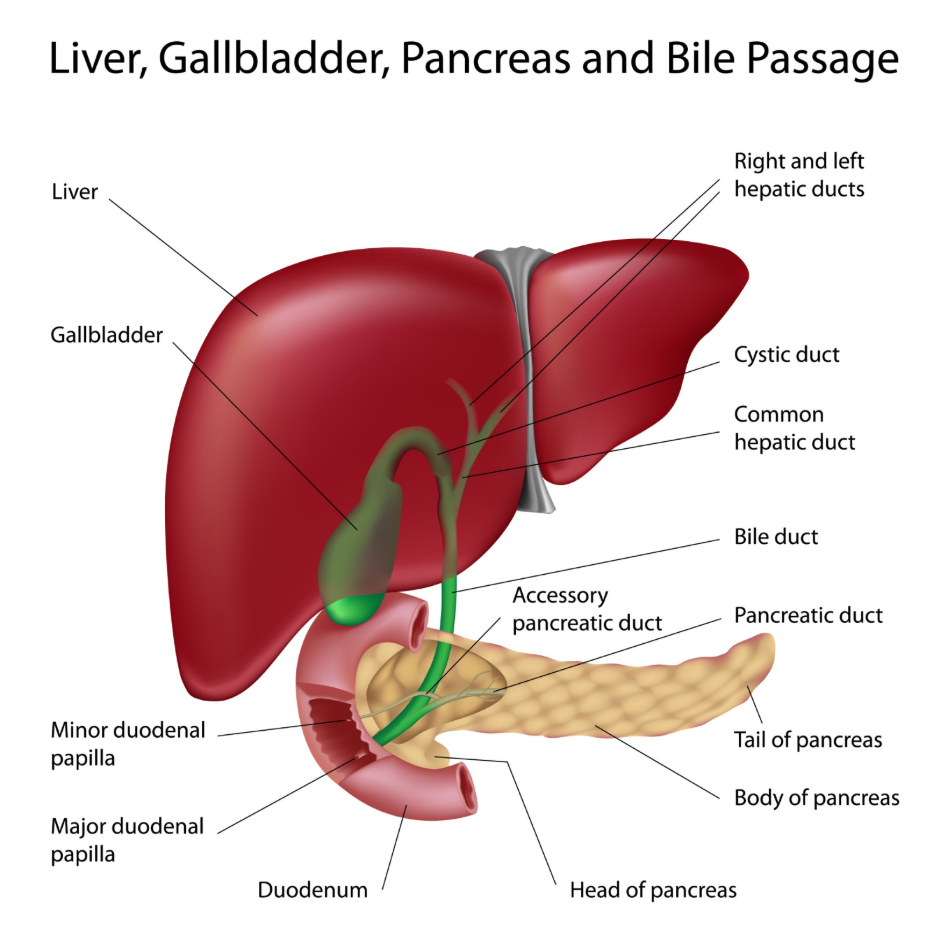
Computed Tomography (CT) scans or Magnetic Resonance Imaging (MRI) may be used to provide more detailed images if the ultrasound results are inconclusive. These scans can identify the presence of regenerative nodules, which are clusters of liver cells surrounded by scar tissue. The GOV.UK health pages indicate that the use of high-resolution imaging is essential for the early detection of liver disease and for monitoring patients who have progressed to advanced scarring. These tests are also vital for screening for primary liver cancer, which is a known complication of chronic cirrhosis.
Transient Elastography and Liver Stiffness
Transient elastography, commonly known by the brand name FibroScan, is a non-invasive test that has become a standard method for confirming liver cirrhosis in the UK. This technology uses ultrasound pulses to measure the “stiffness” of the liver tissue; because scar tissue is much harder than healthy tissue, the speed at which the pulse travels through the organ provides a direct measurement of fibrosis.
The results are typically given in kilopascals, with higher numbers indicating more severe scarring. NICE clinical guidelines recommend using transient elastography to diagnose cirrhosis in people with chronic liver disease, as it is a reliable and painless alternative to more invasive procedures. This test is particularly useful for identifying “compensated” cirrhosis, where the patient may feel healthy but the liver tissue has already undergone significant structural changes.
The Role of Liver Biopsy
A liver biopsy is an invasive procedure that remains the “gold standard” for confirming cirrhosis when non-invasive tests do not provide a clear answer. During a biopsy, a small needle is inserted through the skin and into the liver to remove a tiny sample of tissue, which is then examined under a microscope by a pathologist. This allows for the direct observation of the scarring patterns and the arrangement of liver cells.
| Test Type | Confirmation Method | Primary Indication |
| FibroScan | Measures tissue stiffness | High stiffness score (kPa) |
| Ultrasound/CT | Visualises organ shape | Shrunken, lumpy, or nodular liver |
| Blood Tests | Measures synthetic function | Low albumin and high clotting time |
| Liver Biopsy | Microscopic analysis | Direct observation of fibrous bands |
While a biopsy provides the most detailed information about the stage and cause of the disease, it is now used less frequently due to the accuracy of modern scans. It is generally reserved for cases where the underlying cause of the liver damage is unknown or when it is necessary to differentiate cirrhosis from other complex liver conditions.
Endoscopy and Portal Hypertension Checks
Once cirrhosis is suspected or confirmed, an endoscopy may be performed to check for internal complications that occur as a result of liver scarring. This procedure involves inserting a thin, flexible tube with a camera down the throat to examine the oesophagus and stomach. The primary goal is to look for varices, which are enlarged, fragile veins caused by the resistance to blood flow through the scarred liver.
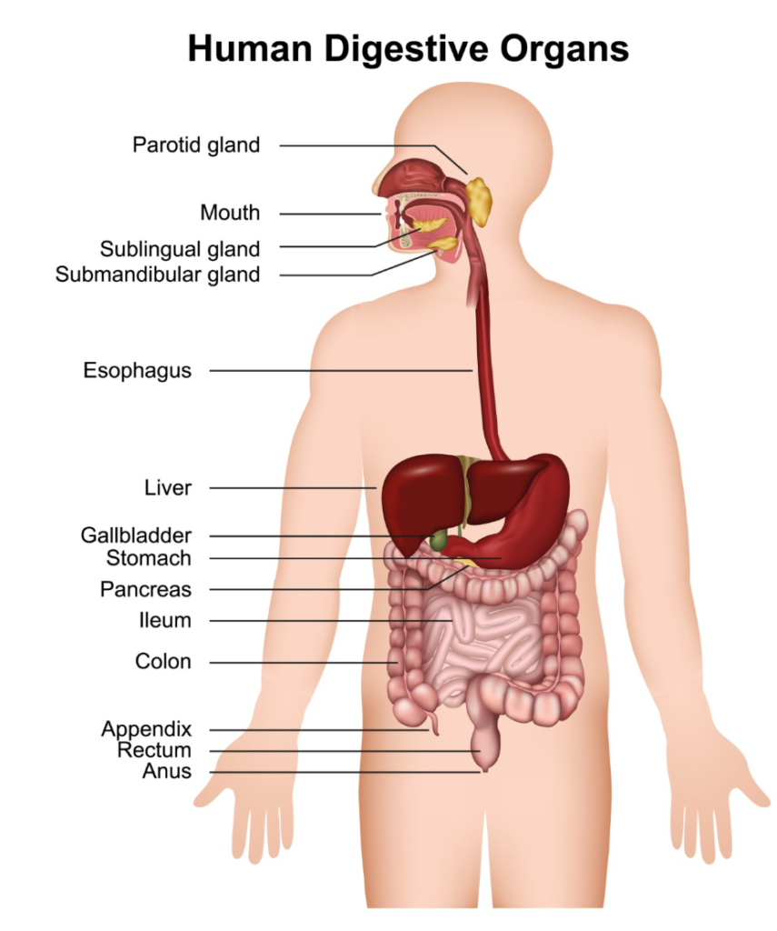
The presence of varices is a strong clinical indicator that cirrhosis has reached a stage where it is affecting the body’s circulatory system. This condition, known as portal hypertension, is one of the most significant risks associated with advanced cirrhosis. Finding and managing these veins early is a critical part of the care plan for any patient with confirmed liver scarring in the UK.
Conclusion
Confirming cirrhosis of the liver requires a tiered diagnostic approach involving blood tests to assess function, imaging to view structural changes, and elastography to measure tissue stiffness. While non-invasive scans are now the primary tool for diagnosis in the UK, a liver biopsy remains available for complex cases where direct tissue analysis is needed. Identifying cirrhosis early through these tests is vital for managing the condition and preventing the transition to liver failure. If you experience severe, sudden, or worsening symptoms, call 999 immediately.
Can a normal ultrasound miss cirrhosis?
In the very early stages of scarring, a standard ultrasound may appear normal, which is why more sensitive tests like a Fibro Scan are often used.
Is the Fibro Scan procedure painful?
No, a Fibro Scan is entirely non-invasive and feels like a slight flick or vibration against the skin over your liver.
How long do blood test results take?
Routine liver function and clotting results are usually available within a few days, though some specialised markers may take longer.
Why do I need an endoscopy if my liver is the problem?
Scarring in the liver blocks blood flow, which can cause veins in your throat to swell; an endoscopy checks for these to prevent internal bleeding.
Does a diagnosis of cirrhosis always require a biopsy?
No, most cases in the UK are now confirmed using a combination of blood tests and specialised scans without the need for a needle biopsy.
What is a “stiffness” score?
The stiffness score measured by a FibroScan indicates how much scar tissue is present; healthy livers are soft, while cirrhotic livers are very stiff.
Can these tests tell what caused the cirrhosis?
Blood tests can identify viruses or genetic markers, and a biopsy can show specific patterns of damage that help identify the cause.
Authority Snapshot (E-E-A-T)
This article provides medically factual health education regarding the diagnosis of cirrhosis, strictly aligned with NHS and NICE clinical protocols. The content is developed by a professional medical writing team and reviewed by Dr. Rebecca Fernandez, a UK-trained physician with experience in internal medicine, surgery, and emergency care. All information follows current UK public health standards to ensure accuracy and patient safety.