The detection of liver cancer in its early stages primarily relies on a structured surveillance programme for individuals with known risk factors, as the disease often develops without causing noticeable symptoms. Because the liver can continue to function effectively even when a small tumour is present, physical signs are rarely a reliable indicator of early malignancy. In the United Kingdom, healthcare professionals use a combination of periodic imaging and blood tests to monitor patients who have conditions such as cirrhosis or chronic viral hepatitis. This proactive approach aims to identify abnormal cellular growths when they are small and localised, which significantly expands the range of effective medical interventions. Understanding the specific diagnostic tools used in the UK helps patients engage with their clinical care and adhere to the regular monitoring schedules necessary for maintaining liver health.
What We’ll Discuss in This Article
- The role of abdominal ultrasound in identifying small liver masses.
- The use of alpha-fetoprotein blood tests as a tumour marker.
- When advanced imaging like CT or MRI scans is required for confirmation.
- The importance of the six-monthly surveillance cycle for high-risk patients.
- How multidisciplinary teams evaluate test results to guide care.
- Rare instances where a liver biopsy may be used for diagnostic clarity.
Abdominal Ultrasound as a Primary Detection Tool
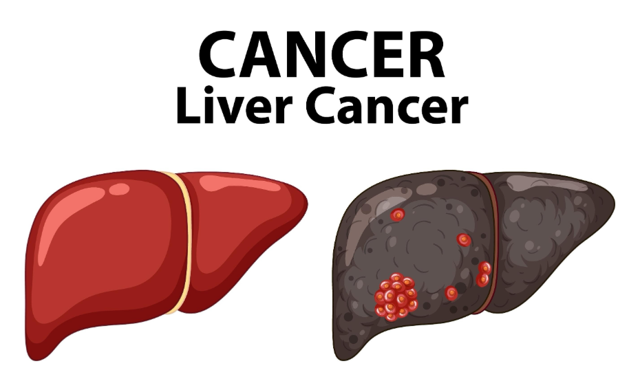
Abdominal ultrasound is the most common non-invasive test used for the early detection of liver cancer because it allows clinicians to visualise the internal structure of the liver without using radiation. During an ultrasound, high-frequency sound waves are bounced off the liver to create a real-time image of the organ, helping sonographers identify any nodules or masses that should not be there. The NHS states that an ultrasound scan is often the first test used to check the liver for tumours and is a standard part of surveillance for people with cirrhosis.
For patients at high risk, an ultrasound is typically performed every six months. This frequency is based on the average growth rate of primary liver cancer cells, ensuring that if a tumour does develop, it is likely to be found while it is still under a specific size. While ultrasound is excellent for screening, its accuracy can sometimes be affected by the presence of extensive scar tissue or the patient’s body composition, which may lead to the use of follow-up tests if an image is unclear.
Alpha-fetoprotein Blood Tests
The alpha-fetoprotein test is a specific blood investigation used alongside imaging to detect potential markers of liver cancer that may not be visible on a scan alone. Alpha-fetoprotein is a protein typically produced by a developing fetus, but its levels can rise significantly in adults when certain types of liver cancer cells are present. In the UK, this test is often used as a supplementary tool to monitor patients with chronic liver conditions.
However, it is important to note that a high alpha-fetoprotein level does not always mean cancer is present, as levels can also rise due to general liver inflammation or pregnancy. Conversely, some early liver cancers do not produce this protein at all. Because of these variations, UK clinicians never use the blood test in isolation; instead, it is interpreted in conjunction with ultrasound findings to provide a more complete picture of the patient’s liver health.
Advanced Imaging: CT and MRI Scans
If an initial ultrasound scan or blood test suggests an abnormality, advanced imaging such as Computed Tomography or Magnetic Resonance Imaging is used to provide a more detailed assessment. These scans offer a much higher resolution and can help specialists determine the exact size, location, and blood supply of a suspected tumour. A CT scan uses X-rays to create cross-sectional “slices” of the liver, while an MRI uses strong magnetic fields to produce detailed internal images.
| Feature | CT Scan | MRI Scan |
| Technology | X-rays and computer processing | Magnetic fields and radio waves |
| Detail Level | High resolution for physical structures | Excellent for soft tissue and blood vessels |
| Contrast | Usually involves an iodine-based dye | Often uses a gadolinium-based contrast |
| Duration | Very quick (minutes) | Longer (30 to 45 minutes) |
NICE clinical guidelines state that if an ultrasound identifies a new nodule in a patient with cirrhosis, a dynamic contrast-enhanced CT or MRI must be performed to confirm whether the growth is malignant. These scans are essential for distinguishing between harmless regenerative nodules caused by cirrhosis and actual cancerous tumours, allowing the medical team to plan the most appropriate course of action.
The Importance of the Surveillance Programme
The effectiveness of early detection in the UK is largely dependent on the patient’s adherence to the six-monthly surveillance programme designed for high-risk groups. This programme is specifically for individuals with a confirmed diagnosis of liver cirrhosis or certain stages of chronic Hepatitis B. Because liver cancer can develop silently, missing even one scheduled scan can allow a tumour to grow to a stage where it is harder to treat.
The GOV.UK health pages indicate that consistent participation in liver surveillance programmes is a key factor in improving long-term outcomes for patients with chronic liver disease. This structured approach ensures that the “silent” nature of the disease is bypassed by regular clinical oversight. For many patients, these routine check-ups provide peace of mind and ensure that any changes in liver health are addressed with the full support of a specialist multidisciplinary team.
Liver Biopsy for Diagnostic Clarity
In rare cases where imaging and blood tests do not provide a definitive answer, a liver biopsy may be considered to confirm the presence of cancerous cells. This procedure involves using a thin needle to remove a small sample of tissue from the suspected tumour, which is then examined under a microscope by a pathologist. A biopsy can provide specific information about the grade of the cancer and its genetic characteristics.
Because the liver is a highly vascular organ and a biopsy carries a small risk of bleeding, it is not used as a first-line detection tool in the UK. Specialists will only recommend a biopsy if the benefits of having a definitive tissue diagnosis outweigh the potential risks of the procedure. In most instances, the combination of high-quality CT or MRI scans and tumour markers is sufficient for clinicians to reach a conclusion without needing a biopsy.
Conclusion
Tests for early liver cancer primarily involve regular ultrasound scans and alpha-fetoprotein blood tests performed every six months for individuals at high risk. If these initial screenings identify an abnormality, advanced CT or MRI scans are used to provide the detailed confirmation required for a medical conclusion. Adhering to this structured surveillance programme is the most effective way to ensure that any cellular changes are caught at the earliest possible stage. Consistent clinical monitoring remains the foundation of liver health management in the United Kingdom.
Why do I need a scan every six months if I feel healthy?
Early liver cancer rarely causes symptoms, so the scan is designed to find tumours while they are still small and before you feel unwell.
Is the ultrasound scan painful?
No, the ultrasound is completely non-invasive and painless; it involves a small amount of gel and a handheld device moved over your skin.
Can a normal blood test find liver cancer?
Standard liver function tests may not detect cancer; a specific test for alpha-fetoprotein is used, but it must be combined with a scan for accuracy.
What happens if my ultrasound shows a small lump?
If a new nodule is found, you will usually be referred for a more detailed CT or MRI scan to determine exactly what the lump is.
Are CT scans and MRI scans safe?
Both are standard medical procedures; your clinical team will review your history to ensure the specific scan and any contrast dye used are appropriate for you.
Does everyone with a fatty liver need cancer screening?
No, screening is usually reserved for those who have developed significant scarring (cirrhosis) or have specific types of chronic viral hepatitis.
How long does it take to get the results of these tests?
Ultrasound results are often available quickly, while more complex CT or MRI scans and blood markers may take a few days to be fully reviewed by specialists.
Authority Snapshot (E-E-A-T)
This article provides medically factual health education regarding the detection of liver cancer, strictly aligned with NHS and NICE clinical guidelines. The content is developed by a professional medical writing team and reviewed by Dr. Rebecca Fernandez, a UK-trained physician with experience in internal medicine, surgery, and emergency care. All information follows current UK public health protocols to ensure accuracy and patient safety.