




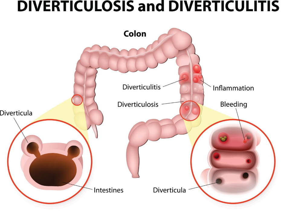
Diverticulitis is typically diagnosed during a flare-up using a combination of physical examinations, blood tests, and diagnostic imaging, with Computed Tomography (CT) scans considered the gold standard for confirmation. When the small pouches in the colon wall, known as diverticula, become inflamed or infected, they cause acute symptoms that require prompt clinical assessment to ensure appropriate management. In the United Kingdom, healthcare professionals follow structured protocols to distinguish diverticulitis from other causes of abdominal pain, such as appendicitis or inflammatory bowel disease. The diagnostic process focuses on identifying the location and severity of the inflammation, as well as checking for potential complications like abscesses or perforations. Because an active flare-up makes the bowel wall fragile, certain invasive tests are avoided until the inflammation has subsided. Understanding the sequence of these tests helps patients navigate their care pathway during what can be a painful and concerning episode. By utilising evidence-based diagnostic tools, the UK healthcare system ensures that patients receive targeted treatment to resolve the infection and protect the structural integrity of the large intestine.
What We’ll Discuss in This Article
- The role of clinical assessment and physical examination during a flare-up.
- Why blood tests are essential for measuring infection and inflammation.
- The importance of CT scans in confirming the presence of diverticulitis.
- Why colonoscopies are generally avoided during the acute inflammatory phase.
- Alternative imaging options for specific patient groups.
- UK clinical pathways for staging the severity of the condition.
Clinical Assessment and Physical Examination
The initial diagnosis of diverticulitis begins with a thorough clinical assessment where a healthcare professional evaluates the patient’s symptoms and performs a physical examination of the abdomen. Clinicians look for specific indicators such as localised tenderness, particularly in the lower left quadrant, which is the most common site for diverticular inflammation in the UK. The NHS states that diverticulitis is usually diagnosed by a GP who will ask about your symptoms and examine your tummy.
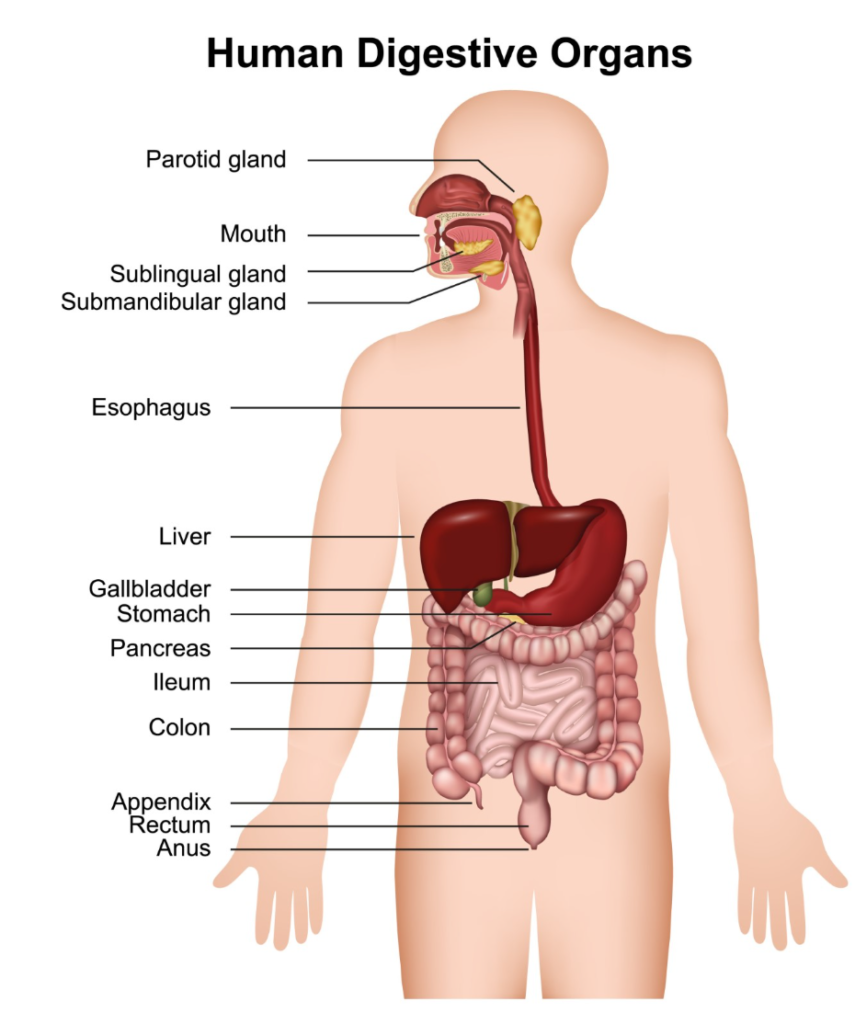
During the examination, the doctor may check for rebound tenderness, a clinical sign where pain increases when pressure is suddenly released, suggesting that the inflammation may be affecting the lining of the abdominal cavity. They will also take a detailed history of the pain’s onset, any changes in bowel habits, and the presence of systemic symptoms like fever or nausea. This initial stage is vital for ruling out other acute surgical conditions. While a physical exam alone cannot confirm diverticulitis, it provides the necessary clinical context to determine which further tests are required to reach a definitive conclusion.
Blood Tests for Infection and Inflammation Markers
Blood tests are used as a secondary diagnostic tool to provide biochemical evidence of an active infection and to measure the severity of the body’s inflammatory response. A Full Blood Count (FBC) is typically performed to check for an elevated white blood cell count, which is a common sign that the immune system is responding to a bacterial infection within the diverticular pouches. NICE clinical guidelines for diverticular disease management recommend using blood tests to look for markers of inflammation, such as C-reactive protein (CRP), to help diagnose acute diverticulitis.
A significantly raised CRP level often correlates with the severity of the flare-up and can help clinicians decide if a patient can be managed at home or requires hospital-based care. These tests also help rule out other issues; for example, liver function tests might be checked if the pain is in the upper abdomen to ensure the gallbladder or liver are not involved. While blood tests are not specific enough to diagnose diverticulitis on their own, they are an essential part of the UK diagnostic pathway, providing a measurable baseline to monitor how well the patient responds to treatment.
Computed Tomography (CT) Scans as the Gold Standard
A CT scan of the abdomen and pelvis is the most reliable test for diagnosing diverticulitis during a flare-up because it can clearly visualise the colon wall, the presence of diverticula, and any surrounding inflammation. During the scan, X-rays are used to create detailed cross-sectional images of the internal organs. In many UK hospitals, a contrast dye may be used to make the structures of the bowel stand out more clearly on the images.
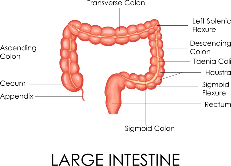
The CT scan allows radiologists to identify thickened bowel walls, “fat stranding” (which indicates inflammation in the surrounding fat), and complications such as abscesses or air bubbles outside the colon that suggest a perforation.
Why Colonoscopies are Avoided During Flare-ups
Invasive visual tests like a colonoscopy are generally avoided during the acute phase of a diverticulitis flare-up because the inflammation makes the colon wall fragile and increases the risk of causing a perforation. A colonoscopy involves inserting a camera into the bowel and inflating it with air to see the lining. During an active infection, the pressure from the air and the movement of the scope could cause a weakened pouch to burst.
The GOV.UK health pages indicate that while a colonoscopy is a vital tool for bowel health, it is usually postponed until at least six to eight weeks after a diverticulitis flare-up has resolved. Once the inflammation has subsided and the tissue has healed, a colonoscopy is often scheduled to confirm the diagnosis and ensure that no other structural issues, such as polyps, were hidden by the previous swelling. This staged approach is a cornerstone of UK gastrointestinal safety protocols, ensuring that the patient’s immediate infection is managed safely before performing a thorough internal inspection.
Ultrasound and Alternative Imaging Options
Ultrasound may be used as an alternative or supplementary diagnostic tool, particularly in cases where a CT scan may not be suitable, such as in pregnant patients or when a quick bedside assessment is needed. An ultrasound uses high-frequency sound waves to create images of the abdominal organs and can often detect the thickened bowel walls and localised fluid collections associated with diverticulitis.
While ultrasound is less definitive than a CT scan, it is non-invasive and does not involve radiation. In some UK clinical settings, if the diagnosis is relatively clear from the physical exam and blood tests, an ultrasound may be used to look for specific complications like a large abscess. However, if the ultrasound results are inconclusive, a CT scan remains the preferred follow-up. The choice of imaging is always tailored to the individual’s clinical needs, ensuring that the most effective and safest tool is used to stage the flare-up and guide the management plan.
UK Clinical Pathways for Staging and Management
The diagnostic pathway in the United Kingdom follows a logical progression from the least invasive to more detailed tests, ensuring that the severity of the diverticulitis is accurately identified. Once the CT scan and blood tests have provided a clear picture, the condition is usually classified as either uncomplicated or complicated. Uncomplicated diverticulitis involves localised inflammation and is often managed with rest and a temporary clear fluid diet.
Complicated diverticulitis involves an abscess, perforation, or obstruction and usually requires admission to a UK hospital for intravenous antibiotics and close monitoring. This staging process is vital because it determines the intensity of the treatment required. Throughout the process, the healthcare team uses the diagnostic results to monitor the patient’s progress. If the inflammatory markers do not decrease or if the pain worsens, the tests may be repeated to ensure no new complications have developed. This integrated approach ensures that every patient receives a level of care appropriate to the specific nature of their diverticulitis flare-up.
Conclusion
Diagnosing diverticulitis during a flare-up in the UK involves a combination of physical examination, blood tests for inflammatory markers, and a CT scan to confirm the localised infection. While the CT scan is the most effective tool for staging the severity and identifying complications, invasive tests like colonoscopies are avoided until the inflammation has resolved. This staged diagnostic pathway ensures that the immediate infection is managed safely and effectively. Understanding these tests allows patients to participate actively in their recovery and long-term bowel health management. If you experience severe, sudden, or worsening symptoms, call 999 immediately.
Can a stool test diagnose diverticulitis?
Stool tests are not used to diagnose diverticulitis during a flare-up, though they may be used later to rule out other issues like inflammatory bowel disease.
Why can’t I just have a colonoscopy straight away?
During a flare-up, the colon wall is very fragile; a colonoscopy carries a high risk of causing a tear or burst pouch until the inflammation has healed.
Is a CT scan safe if I have had many before?
Your clinical team will only order a CT scan if the benefits of an accurate diagnosis outweigh the risks of radiation exposure.
How long do blood test results take?
In most UK urgent care settings, basic blood test results for inflammation and infection markers are usually available within a few hours.
Will a regular X-ray find diverticulitis?
A standard X-ray cannot see the inflammation of the pouches, but it might be used to check for free air in the abdomen if a burst bowel is suspected.
Do I need to go to the hospital for these tests?
Initial assessments often happen at a GP surgery, but a CT scan and detailed blood work usually require a visit to a hospital or diagnostic centre.
What if my tests are all normal but I still have pain?
If tests do not show diverticulitis, the clinical team will investigate other potential causes of your symptoms, such as IBS or muscular issues.
Authority Snapshot (E-E-A-T)
This article provides medically factual health education regarding the tests used to diagnose diverticulitis, strictly aligned with NHS and NICE clinical guidelines. The content is developed by a professional medical writing team and reviewed by Dr. Rebecca Fernandez, a UK-trained physician with extensive experience in internal medicine, emergency care, and general surgery. All information follows current UK public health protocols to ensure clinical accuracy and patient safety.



