




Basal cell carcinoma, often referred to as a rodent ulcer, is the most common form of skin cancer in the United Kingdom. While it rarely spreads to other parts of the body, it can cause significant local damage if left untreated, particularly on the delicate tissues of the face and neck. Because these areas receive the highest amount of cumulative sun exposure, they are the most frequent sites for these lesions to develop. Recognising the early physical signs of a basal cell carcinoma is essential for ensuring that treatment is straightforward and results in minimal scarring.
What We’ll Discuss in This Article
- The classic appearance of nodular basal cell carcinoma
- Identifying the pearly border and telangiectasia in early lesions
- How a rodent ulcer develops through central ulceration
- Recognising the flatter, scaly appearance of superficial BCC
- Distinguishing basal cell carcinoma from benign skin spots
- Common locations on the face and neck for these lesions
- Why a slow growth rate can lead to a delayed diagnosis
The classic appearance of nodular basal cell carcinoma
The most frequent type of basal cell carcinoma found on the face and neck is the nodular variety. This typically starts as a small, firm, and raised bump on the skin surface. Unlike a common spot or blemish, it does not resolve over time but slowly increases in size over several months or even years. These nodules are often described as being skin-coloured, pink, or slightly translucent, giving them a distinctive appearance that stands out from the surrounding healthy tissue.
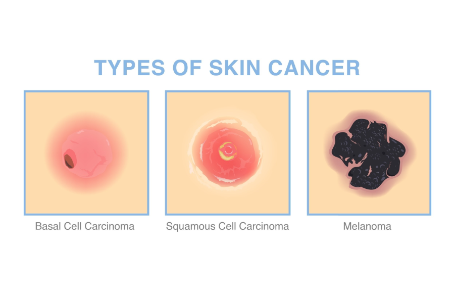
The National Health Service describes a typical basal cell carcinoma as a small, slow-growing, shiny pink or pearly white lump with a translucent or waxy appearance. Because they grow so slowly, many people mistake them for a persistent pimple or a harmless age-related skin change. However, a key clinical feature of a basal cell carcinoma is its inability to heal; even if it appears to scab over, the underlying lump remains and will eventually begin to grow again.
Identifying the pearly border and surface blood vessels
When examining a suspected basal cell carcinoma under a bright light, clinicians look for two specific diagnostic features: a pearly border and telangiectasia. The pearly border refers to the shiny, rolled edge of the lesion that often catches the light. This gives the growth a slightly crater-like appearance as it matures. Telangiectasia are tiny, thread-like red blood vessels that become visible just beneath the surface of the translucent skin within the nodule.
These fine blood vessels are a result of the tumour creating its own blood supply as it expands. While they may be difficult to see with the naked eye, they are very clear when viewed through a dermatoscope by a dermatologist. The presence of these vessels, combined with the waxy or pearly sheen of the skin, is a strong indicator of a basal cell carcinoma. If you notice a persistent bump on your face that has these fine red lines on its surface, it warrants a professional medical review.
The development of a rodent ulcer through ulceration
As a nodular basal cell carcinoma grows, the centre of the lump may break down to form an open sore or ulcer. This is why the condition was historically called a rodent ulcer, as it appears to slowly eat away at the local tissue. The ulcer often crusts or bleeds, especially after minor trauma like washing the face or drying with a towel. It may seem to heal for a few days before the scab falls off and the bleeding returns.
This cycle of bleeding, scabbing, and failing to heal is a hallmark sign of a basal cell carcinoma. While it is rarely painful, the persistent nature of the sore is a cause for concern. On the face and neck, these ulcers can grow downwards into the deeper layers of the skin and, if neglected, can eventually affect the underlying cartilage or bone. Early surgical intervention at this stage is highly effective and prevents the need for more complex reconstructive surgery later on.
Recognising superficial and morphoeic BCC types
While the nodular type is the most common on the face, other forms of basal cell carcinoma can also appear. Superficial basal cell carcinoma often looks like a flat, scaly red patch on the skin. It can be mistaken for a patch of eczema or psoriasis, but unlike those conditions, it does not respond to moisturising creams or steroid ointments. It often has a very fine, shiny thread-like border if you look closely under good lighting.
A rarer and more difficult type to spot is the morphoeic or sclerosing basal cell carcinoma. This often looks like a firm, waxy scar or an area of thickened skin that has appeared without any previous injury. The borders are often poorly defined, making it hard to tell where the lesion ends and healthy skin begins. Because it can look so much like a normal scar, it is often diagnosed later than other types. Any scar-like area on your face that is enlarging or changing should be assessed by a specialist.
Distinguishing BCC from benign skin spots
It is often challenging for the general public to tell the difference between a basal cell carcinoma and various harmless skin spots. Common conditions that can mimic a basal cell carcinoma include fibrous papules, which are small skin-coloured bumps often found on the nose, and sebaceous hyperplasia, which are yellowish, doughnut-shaped bumps caused by enlarged oil glands. Unlike a basal cell carcinoma, these benign spots typically remain stable in size and do not bleed or ulcerate.
Clinical guidelines from NICE emphasize that any new or changing skin lesion that persists for more than four weeks should be reviewed by a General Practitioner for a potential referral. A dermatologist can often make a definitive diagnosis using a dermatoscope without the need for an immediate biopsy. If there is any doubt, a small sample of the skin will be taken under local anaesthetic to confirm the nature of the growth.
Conclusion
A typical basal cell carcinoma on the face or neck appears as a slow-growing, pearly, or waxy lump that may have visible blood vessels on its surface. It often follows a cycle of scabbing and bleeding, leading to the formation of a rodent ulcer if left untreated. Recognising these persistent, non-healing sores early is the key to simple and effective treatment with minimal cosmetic impact.
If you experience severe, sudden, or worsening symptoms, call 999 immediately.
Does a basal cell carcinoma ever itch or hurt?
Most basal cell carcinomas are completely painless and do not itch, which is often why people wait a long time before seeing a doctor.
How fast do these skin cancers grow?
They grow very slowly, often taking several months or even years to increase in size by just a few millimetres.
Are basal cell carcinomas always pink or red?
No, they can be skin-coloured, white, waxy, or even brown or black in people with darker skin tones.
Will a basal cell carcinoma go away on its own?
No, a basal cell carcinoma will not disappear without treatment and will continue to grow slowly into the surrounding skin.
Can I get a basal cell carcinoma on my scalp?
Yes, the scalp is a common site, especially for individuals with thinning hair who receive significant sun exposure in that area.
Is a basal cell carcinoma caused by a one-off sunburn?
While sunburns contribute, basal cell carcinomas are more commonly linked to the cumulative sun exposure received over many years of life.
What is the best way to prevent these cancers on my face?
Daily use of a high-factor sunscreen and wearing a wide-brimmed hat during the summer months are the most effective preventative measures.
Authority Snapshot (E-E-A-T)
This article is designed to provide clear and factual information regarding skin health for the general public. The content is written by the Medical Content Team and has been reviewed by Dr. Stefan Petrov. He is a UK-trained physician with an MBBS and postgraduate certifications including Basic Life Support (BLS), Advanced Cardiac Life Support (ACLS), and the UK Medical Licensing Assessment (PLAB 1 & 2). He has hands-on experience in general medicine, surgery, anaesthesia, ophthalmology, and emergency care. Dr. Petrov has worked in both hospital wards and intensive care units, performing diagnostic and therapeutic procedures, and has contributed to medical education by creating patient-focused health content and teaching clinical skills to junior doctors.



