What tests are used to diagnose coronary artery disease in the first place?
Diagnosing coronary artery disease is rarely done with a single test. Instead, doctors use a combination of checks to build a picture of your heart’s health. The process usually starts with simple, non-invasive tests to assess your risk factors and heart rhythm, followed by more advanced imaging to look directly at the arteries. Understanding this pathway can reduce anxiety and help you prepare for your appointments.
What We’ll Discuss in This Article
- The initial tests performed by a GP (ECG and bloods).
- The difference between a resting ECG and a stress test.
- How advanced imaging like CT Coronary Angiograms works.
- The role of invasive angiography as a ‘gold standard.’
- What blood tests tell us about heart risk (Troponin and Lipids).
- Differentiating between functional tests and anatomical scans.
- When to expect a referral to a cardiologist.
What are the first tests used to check for heart disease?
The first tests usually involve a physical exam, blood pressure check, and blood tests to measure cholesterol (lipid profile) and blood sugar (HbA1c). A resting Electrocardiogram (ECG) is almost always performed to check the heart’s electrical rhythm and look for signs of previous heart attacks or strain.
Initial GP Assessment
Before sending you to a specialist, a GP will assess your ‘pre-test probability’ of having CAD.
- Blood Tests: These check for risk factors like high cholesterol and diabetes. They also check kidney function, as this affects what medication or scans you can have.
- Resting ECG: The NHS states that an electrocardiogram (ECG) is a simple test that can be used to check your heart’s rhythm and electrical activity. It involves sticking sensors to your skin. While it can detect old heart attacks, a normal resting ECG does not rule out coronary artery disease.
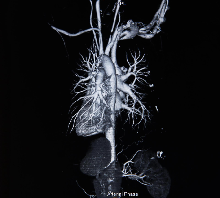
How do doctors see the blocked arteries?
To see the actual blockages, doctors typically use a CT Coronary Angiogram (CTCA) or an Invasive Coronary Angiogram. A CTCA is a specialised X-ray scan that creates 3D images of the heart vessels, while an invasive angiogram involves inserting a catheter into the wrist or groin to inject dye directly into the arteries.
Shutterstock
Imaging Options
- CT Coronary Angiogram: This is now the first-line test for most people with stable chest pain. It is non-invasive, quick, and excellent at ruling out disease.
- Invasive Angiogram: This is the ‘gold standard.’ It is invasive but allows the doctor to treat the blockage (with a stent) at the same time if needed.
- Echocardiogram: An ultrasound scan that looks at the structure and pumping function of the heart, rather than the arteries themselves.
Why are these tests ordered? (The Underlying Cause)
These tests are ordered to detect atherosclerosis, the buildup of fatty plaque in the arteries, and to assess if this buildup is limiting blood flow (ischaemia). Functional tests like a perfusion scan or stress echo specifically look for areas of the heart muscle that are starving of oxygen under stress.
- Anatomical Tests (CT/Angiogram): Show what the blockage looks like (e.g., ‘70% narrowing’).
- Functional Tests (Stress Echo/MRI): Show how the blockage affects blood flow (e.g., ‘muscle wall stops moving during exercise’).
Triggers for Testing: When should you be checked?
You will typically be referred for these tests if you present with symptoms of angina, such as chest pain triggered by exertion, breathlessness, or palpitations. Doctors use risk scoring systems (like QRISK3) and clinical judgment to decide if your symptoms warrant urgent investigation (Rapid Access Chest Pain Clinic) or routine screening.
- Stable Symptoms: Chest tightness when walking uphill that goes away when you stop.
- Risk Factors: High blood pressure, smoking history, or a family history of premature heart disease may lower the threshold for testing.
- Abnormal Screening: If a routine health check finds an irregular pulse or very high cholesterol, further cardiac testing may be triggered.
Differentiation: CT Angiogram vs. Invasive Angiogram
It is important to understand the difference between these two key tests. A CT Angiogram is a diagnostic scan used to rule out disease, while an Invasive Angiogram is often used to confirm and treat significant disease found on other tests.
| Feature | CT Coronary Angiogram (CTCA) | Invasive Coronary Angiogram |
| Method | X-ray scanner (doughnut shape) | Catheter (tube) in wrist/groin |
| Invasiveness | Minimally (cannula in arm) | Invasive (arterial puncture) |
| Purpose | Diagnosis / Screening | Diagnosis + Treatment (Stent) |
| Recovery | Immediate | Requires hours of bed rest |
| Radiation | Yes | Yes (Fluoroscopy) |
Conclusion
Diagnosing coronary artery disease is a step-by-step process. It typically begins with blood tests and an ECG at your GP, followed by a CT Coronary Angiogram to visualise the arteries. Understanding which test looks for ‘structure’ (anatomy) versus ‘function’ (blood flow) helps explain why you might need more than one type of scan. These tests are vital for catching the disease early and planning the right treatment.
If you are waiting for a test and your chest pain becomes severe, occurs at rest, or lasts more than 15 minutes, do not wait for your appointment. Call 999 immediately.
Can a normal ECG rule out a heart attack?
No. A resting ECG can be completely normal even if you have significant coronary artery disease or are having a ‘Non-STEMI’ heart attack. Further blood tests (troponin) are needed.
Is a stress test the same as an angiogram?
No. A stress test (like running on a treadmill) measures how your heart behaves under load. An angiogram takes a picture of the arteries.
Does a CT angiogram hurt?
No. It is painless, though you will need a cannula (needle) in your arm for the dye, which can give you a warm, flushing sensation.
How long does a heart check-up take?
An initial assessment (ECG and bloods) takes 15–20 minutes. A CT angiogram appointment usually lasts about an hour, though the scan itself takes minutes.
What is a Troponin test?
Troponin is a protein released into the bloodstream when the heart muscle is damaged. High levels usually indicate a recent heart attack.
Can I eat before these tests?
For blood tests (lipids/glucose), you may need to fast. For a CT angiogram, you are often asked to avoid caffeine (which speeds up the heart) for 12 hours beforehand.
Why did the doctor listen to my neck?
They are checking for a ‘bruit’ (a whooshing sound) in the carotid arteries. This can indicate narrowing of the arteries in the neck, which suggests you likely have narrowing in the heart arteries too.
Authority Snapshot
This article was written by Dr. Rebecca Fernandez, a UK-trained physician (MBBS) with extensive experience in cardiology and internal medicine. Having assessed thousands of patients with chest pain in both emergency and outpatient settings, Dr. Fernandez explains the diagnostic pathway for coronary artery disease (CAD). This content has been reviewed to ensure alignment with NHS and NICE protocols, guiding you through what to expect during your heart assessment.
Internal Link Suggestions
- ‘what is an ecg test’
- ‘preparing for an angiogram’
- ‘understanding troponin levels’
- ‘stress echocardiogram explained’
