The specific type of a brain tumour is one of the most significant factors in determining the long-term prognosis and the likely effectiveness of medical treatment. In the United Kingdom, healthcare professionals categorise tumours by the cells they originate from and their biological behaviour to establish a clear clinical outlook. While some types are slow-growing and carry a more positive prognosis, others are infiltrative and require more intensive management. By adhering to the evidence-based standards of the NHS and NICE, multidisciplinary teams use this classification to tailor care plans to the individual. Understanding how different tumour types behave is essential for patients navigating the complexities of neurological health. This article explores the relationship between tumour classification and prognosis, the role of tumour grading, and the integrated support provided within the UK healthcare framework.
What We’ll Discuss in This Article
- The distinction between primary and secondary brain tumours.
- How the cell of origin influences the speed of tumour growth.
- The role of the World Health Organisation grading system in prognosis.
- The impact of common tumour types such as gliomas and meningiomas.
- Why molecular and genetic markers are vital for modern outlooks.
- How UK clinical teams coordinate long-term surveillance and monitoring.
Primary and Secondary Tumour Classifications
A fundamental factor in determining prognosis is whether the brain tumour is primary, meaning it started in the brain, or secondary, meaning it spread from another part of the body. Primary tumours are classified based on the type of brain cell they involve, such as glial cells or meningeal tissue, and their outlook depends heavily on their grade. The NHS states that a primary brain tumour is one that starts in the brain, while a secondary brain tumour is cancer that has spread to the brain from elsewhere.
Secondary tumours, also known as brain metastases, generally indicate that the original cancer is at an advanced stage, which significantly influences the overall clinical prognosis. In the United Kingdom, specialists manage primary and secondary tumours through different clinical pathways. For primary tumours, the focus is often on local control through surgery or radiation. For secondary tumours, the management involves treating both the brain lesion and the primary cancer site. This distinction is the first step used by the multidisciplinary team to establish a realistic expectation for the patient health journey and to decide on the most appropriate interventions.
The Role of Cell Origin in Growth Patterns
The type of cell from which a tumour grows determines its “behavioural profile,” including how likely it is to invade surrounding healthy brain tissue. Gliomas, which develop from the glial cells that support nerve cells, are often infiltrative, meaning they do not have clear borders and can be more difficult to treat completely.
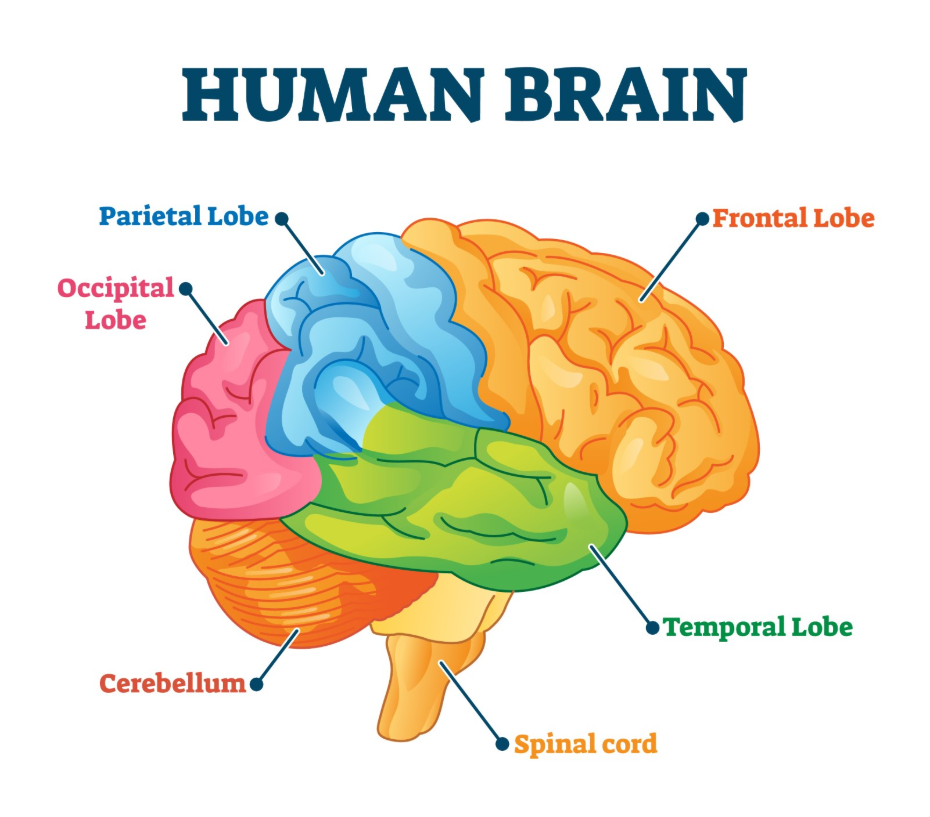
Conversely, tumours like meningiomas grow from the protective membranes surrounding the brain and often have distinct margins.
| Tumour Type | Originating Cells | Typical Growth Pattern |
| Glioma | Glial cells (supporting cells). | Often infiltrative and lacks clear margins. |
| Meningioma | Meninges (protective membranes). | Usually slow-growing and well-defined. |
| Acoustic Neuroma | Vestibular nerve (hearing/balance). | Benign and usually very slow-growing. |
| Pituitary Tumour | Pituitary gland (hormone centre). | Often benign but can affect hormone levels. |
In the United Kingdom, the pathology report identifying the cell type is vital for predicting how a tumour will respond to surgery. A well-defined meningioma often carries a more positive prognosis because a full surgical removal is more likely. Infiltrative gliomas, however, typically require a combination of surgery and adjuvant therapies to manage the microscopic cells that remain. By identifying these growth patterns early, UK clinicians can provide a more accurate prognosis and ensure that the patient receives the necessary level of specialist follow-up.
Influence of the WHO Grading System
The World Health Organisation grading system is used in the United Kingdom to provide a standardised measure of how aggressive a tumour type is, ranging from Grade 1 to Grade 4. NICE clinical guidelines for brain tumours indicate that the grade of the tumour is a primary factor in determining the urgency of treatment and the frequency of follow-up imaging. Grade 1 tumours are slow-growing and often potentially curable, while Grade 4 tumours are highly aggressive and carry a more challenging prognosis.
The grade of a tumour is determined by looking at the cells under a microscope to see how much they differ from normal cells and how quickly they appear to be dividing. A low-grade tumour type can occasionally progress to a higher grade over time, a factor that UK specialists monitor for during long-term surveillance. The prognosis for a Grade 2 glioma is generally more favourable than for a Grade 4 glioblastoma, but the clinical team remains vigilant in both cases. This grading framework ensures that the NHS provides a consistent level of care that is proportionate to the biological threat posed by the specific tumour type.
Prognosis for Common Primary Tumour Types
The prognosis for specific primary brain tumours varies widely across the UK population, with some types being managed successfully over many decades. Meningiomas are the most common primary brain tumour in adults and, because they are often slow-growing and Grade 1, many patients have a normal life expectancy after treatment. In contrast, glioblastomas are more aggressive and require immediate, intensive care to manage the disease.
Other tumour types with distinct outlooks in the UK include:
- Ependymomas: These can occur in children or adults and their prognosis depends on their location within the brain or spinal cord.
- Medulloblastomas: Primarily found in children, these require intensive combined therapy but often have a positive long-term outlook with modern care.
- Lymphomas of the Brain: These respond differently than other tumours and are often managed primarily with chemotherapy.
- Low-Grade Astrocytomas: These often allow for many years of stable health before further treatment is required.
Identifying the exact type of tumour is essential because it allows the medical team to give the patient a clearer understanding of what to expect. In the United Kingdom, the 28-day faster diagnosis target ensures that these details are established quickly so that the appropriate management can begin. By understanding the typical “path” of a specific tumour type, patients and families can better prepare for the practical and emotional aspects of the clinical journey.
Molecular Markers and Genomic Influences
In the United Kingdom, modern prognosis is increasingly determined by molecular markers and genetic signatures found within the tumour cells. These markers provide a deeper level of detail than traditional cell grading alone and can predict how well a patient might respond to chemotherapy or targeted treatments. The GOV.UK health pages provide clinical profiles indicating that genomic testing is now a mandatory part of the diagnostic pathway for many brain tumours in the UK.
Key molecular factors that influence prognosis include:
- IDH Mutation: This marker is often associated with a better prognosis in certain types of gliomas.
- 1p/19q Codeletion: A genetic change that suggests a tumour may be more sensitive to specific treatments.
- MGMT Methylation: Indicates that a tumour is more likely to respond to temozolomide chemotherapy.
- BRAF Mutations: Found in some rare tumours and can be targeted with new, specialised medications.
These markers are identified through specialist laboratory testing of the tumour tissue obtained during a biopsy or surgery. In the UK, the multidisciplinary team integrates these findings into the final prognosis. For example, two patients with the same “type” of glioma may have different outlooks based on their molecular markers. This shift toward “personalised medicine” in the NHS allows for more refined care plans that prioritise treatments with the highest likelihood of success for the individual.
Integrated Monitoring and Follow-up Care
Regardless of the tumour type, every patient in the United Kingdom follows an integrated monitoring pathway to manage their long-term health and to detect any changes in their condition early. The frequency of MRI scans and specialist consultations is determined by the tumour type, its grade, and the response to initial treatment. This “safety netting” is a vital part of UK clinical practice, ensuring that the management plan adapts to the patient’s evolving needs.
The UK follow-up framework involves:
- Regular Neuroimaging: Scheduled scans to check for stability or regrowth of the tumour.
- Clinical Assessment: Regular reviews with a specialist to check physical and cognitive function.
- Supportive Services: Access to neuro-rehabilitation, specialist nurses, and psychological support.
- Coordinated Review: Re-discussion of the case by the multidisciplinary team if the clinical picture changes.
This structured system provides a consistent level of care across the NHS and ensures that patients are not left to manage their condition alone after the initial treatment phase. For those with slow-growing tumour types, the focus may be on maintaining quality of life and functional independence over many years. For more aggressive types, the emphasis remains on close vigilance and prompt intervention if the tumour shows signs of change. This longitudinal approach acknowledges that a brain tumour is a long-term health journey requiring consistent medical oversight.
Conclusion
The type of brain tumour is a primary factor that affects prognosis, influencing how the growth behaves and how it responds to medical interventions. In the UK, the NHS uses a combination of cell origin, WHO grading, and molecular markers to establish a clear clinical outlook for every patient. While low-grade or well-defined tumours often carry a more positive long-term prognosis, infiltrative and high-grade tumours require intensive, coordinated care. Advanced genomic testing is now an essential part of the UK diagnostic pathway, allowing for a more personalised understanding of each case. Following a structured monitoring plan with a multidisciplinary team is vital for managing neurological health over time. If you experience severe, sudden, or worsening symptoms, call 999 immediately.
Can a benign tumour type still be serious?
Yes; even a non-cancerous tumour can be serious if its location puts pressure on vital areas of the brain or if it is difficult to reach surgically.
What is the difference between a grade and a type?
The “type” tells you which cells the tumour started from, while the “grade” tells you how aggressively those cells are dividing and growing.
Does my prognosis change after surgery?
Yes; once the surgeon sees how much of the tumour can be removed and the pathologist provides the final grade, your medical team will refine your outlook.
Why do molecular markers matter for my prognosis?
They provide details about the “genetics” of the tumour, which can tell doctors if the cells are likely to be killed easily by specific drugs.
Can a slow-growing tumour type become aggressive later?
Some low-grade tumours have the potential to change into a higher grade over time, which is why the UK healthcare system uses long-term monitoring.
Is the prognosis for secondary brain tumours the same as primary ones?
No; the prognosis for secondary tumours depends heavily on the type of original cancer and how well it is being controlled in the rest of the body.
Where can I find survival statistics for my specific tumour type?
Your specialist consultant is the best person to discuss this with, as they can explain national statistics in the context of your individual diagnostic markers.
Authority Snapshot (E-E-A-T)
This article provides medically factual health education regarding the impact of tumour type on prognosis, strictly aligned with NHS and NICE clinical guidelines. The content is developed by a professional medical writing team and reviewed by Dr. Rebecca Fernandez, a UK-trained physician with extensive experience in general surgery, cardiology, emergency medicine, and psychiatry. All information follows current UK public health protocols to ensure clinical accuracy and patient safety.
