




The appearance of unexplained red or white patches within the oral cavity is one of the most significant early indicators of potential malignancy or precancerous changes. These patches, clinically referred to as leukoplakia and erythroplakia, represent alterations in the normal cellular structure of the mouth’s lining. While many such changes are benign or related to common infections, their persistence can signal that the squamous cells are undergoing mutations. Identifying these visual changes during a self-examination or a routine dental check-up is a vital step in the early detection and management of oral cancer.
What We’ll Discuss in This Article
- The clinical definitions of leukoplakia and erythroplakia.
- How to distinguish between common oral conditions and concerning patches.
- The biological process of dysplasia and its link to cancer.
- Vulnerable areas in the mouth where these patches frequently appear.
- The importance of the “three-week rule” for monitoring tissue changes.
- What to expect during a professional clinical assessment of oral lesions.
Understanding White Patches (Leukoplakia)
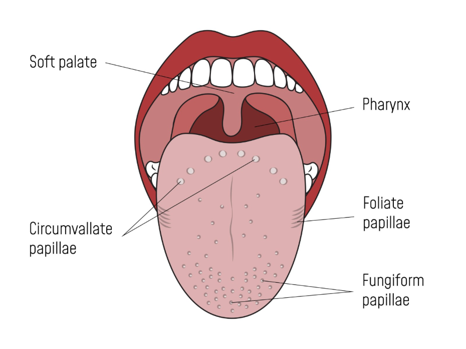
Leukoplakia is a clinical term used to describe a white or greyish patch in the mouth that cannot be wiped or scraped away. These patches develop when the body produces excess keratin (the protein found in hair and nails) in response to chronic irritation or cellular damage. The NHS states that while most cases of leukoplakia are not cancerous, about 1 in 10 cases may eventually lead to oral cancer if left unmonitored. These patches can feel slightly thick, rough, or “leathery” to the touch and are commonly found on the tongue, the floor of the mouth, or the inner lining of the cheeks.
Understanding Red Patches (Erythroplakia)
Red patches, known as erythroplakia, are generally considered more concerning than white patches because they have a higher statistical probability of being cancerous or precancerous at the time of discovery. Erythroplakia appears as a bright red, velvety area that is often clearly defined against the surrounding healthy tissue. These patches occur because the surface layer of the oral lining has become very thin, allowing the underlying blood vessels to show through more prominently. NICE clinical guidelines categorise unexplained erythroplakia as a high-risk symptom that requires an urgent referral to a specialist for investigation.
The Biological Process of Dysplasia
When a clinician observes a red or white patch, they are looking for signs of dysplasia, which refers to the presence of abnormal cells within the tissue. Dysplasia is not cancer itself, but it is a precancerous state where the cells have begun to grow in a disordered way. If the underlying cause—such as chronic tobacco use or alcohol irritation—continues, these dysplastic cells can accumulate further genetic mutations and eventually become invasive cancer cells. Identifying these changes while they are still in a precancerous state offers the highest chance of preventing a full malignancy from developing.
Vulnerable Areas for Tissue Changes
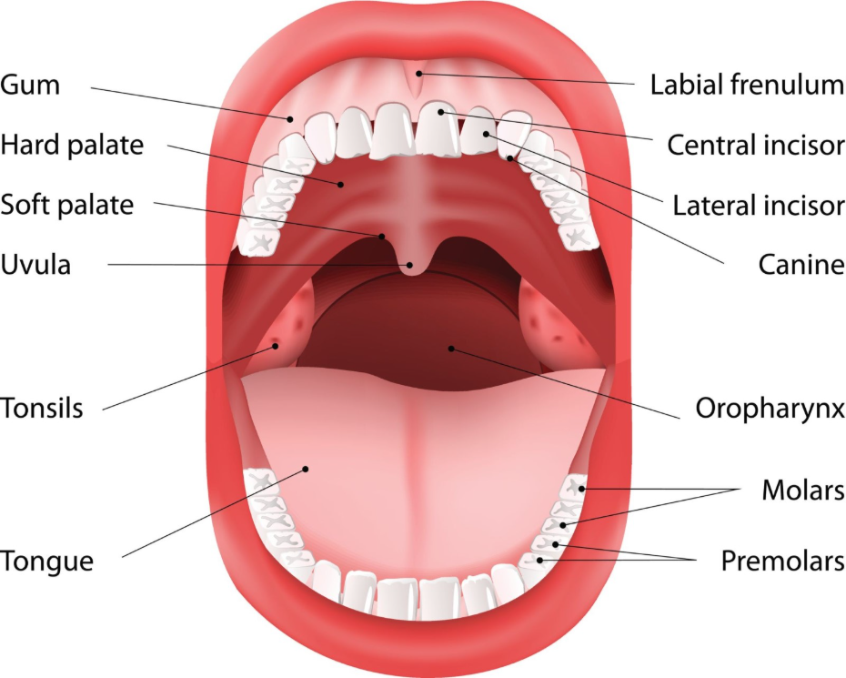
While red and white patches can appear anywhere in the oral cavity, certain locations are associated with a higher risk of malignancy. The floor of the mouth (under the tongue) and the lateral borders (sides) of the tongue are the most critical areas to monitor. These tissues are thinner and more permeable than other parts of the mouth, making them more susceptible to damage from carcinogens. Other areas that require regular inspection include the gums, the roof of the mouth (hard and soft palate), and the inner lining of the cheeks, especially in areas where tobacco or alcohol might make frequent contact.
Distinguishing Cancerous Patches from Benign Conditions
It is important to remember that not all patches in the mouth are related to cancer. Several common conditions can mimic the appearance of leukoplakia or erythroplakia, including:
- Oral Thrush: A fungal infection that creates white patches that can usually be wiped away, leaving a red, sore area underneath.
- Friction Keratosis: White patches caused by a sharp tooth or ill-fitting dentures rubbing against the cheek.
- Lichen Planus: An inflammatory condition that often creates a “lace-like” pattern of white lines on the inner cheeks.
- Geographic Tongue: A benign condition where red, map-like patches appear on the tongue and shift location over time.
Because these conditions can look similar to the early stages of cancer, any patch that does not have a clear cause and does not resolve on its own requires professional evaluation.
When to Seek Professional Advice
The definitive factor for assessing any oral patch is its persistence. If a red or white patch is found in the mouth and does not disappear within three weeks, it is essential to arrange a check-up with a dentist or a GP. Professionals use a combination of visual inspection and, if necessary, a biopsy to determine the exact nature of the tissue change. A biopsy involves taking a small sample of the affected area for laboratory analysis, which is the only way to confirm whether dysplasia or cancer is present. Early detection through this process is the most effective tool for managing oral health.
Conclusion
White and red patches in the mouth are significant early warning signs that should never be ignored if they persist. While leukoplakia is often a response to irritation, erythroplakia carries a higher risk of malignancy and requires immediate clinical attention. By maintaining a regular habit of self-monitoring and following the three-week rule for any tissue changes, individuals can ensure that they are taking a proactive approach to preventing and managing oral cancer.
If you experience severe, sudden, or worsening symptoms, call 999 immediately.
Can I scrape off a leukoplakia patch with a toothbrush?
No, a true leucoplakia patch is firmly attached to the tissue and cannot be removed by scraping or brushing.
Is erythroplakia usually painful?
Erythroplakia can sometimes be tender or have a slight burning sensation, but it is often painless in its early stages.
Can smoking cause white patches in the mouth?
Yes, chronic smoking is a leading cause of leukoplakia as the tissues produce excess keratin to protect themselves from chemical irritation.
What is the difference between oral thrush and oral cancer?
Oral thrush patches can usually be wiped away and are often associated with a metallic taste or soreness, whereas cancerous patches are fixed.
Will a red patch turn into cancer?
Not necessarily, but red patches have a high risk of being precancerous, so they must be professionally assessed as soon as possible.
Do red and white patches ever appear together?
Yes, this is known as “speckled leucoplakia” or “erythroleukoplakia,” and it is often considered higher risk than a purely white patch.
Should I stop drinking alcohol if I find a patch?
Reducing alcohol consumption is a positive health step, but you still need a professional diagnosis to identify the cause of the patch.
Authority Snapshot (E-E-A-T)
This article provides evidence-based information on the significance of red and white oral patches to support public health awareness. The content is produced by our Medical Content Team and reviewed by Dr. Stefan Petrov, a UK-trained physician, to ensure clinical accuracy. All information provided is strictly aligned with the safety and diagnostic guidelines of the NHS and NICE.



