




Deep vein thrombosis (DVT) is diagnosed through a structured clinical pathway that integrates physical examinations, probability scoring, and objective medical imaging to confirm the presence of a blood clot. In the United Kingdom, healthcare professionals follow evidence-based protocols to ensure an accurate physiological understanding of the circulatory system while maintaining biological homeostasis and systemic stability. By utilised integrated NHS pathways, individuals receive a stable foundation for health maintenance through specialist reviews focused on achieving long-term stability within a secure medical environment. This coordinated effort prioritises the safety of the individual, providing a secure framework for building long-term health wellbeing through the systematic monitoring of the vascular environment and the use of validated clinical observations within the UK healthcare framework to achieve long-term stability in a professional environment.
What We’ll Discuss in This Article
- The initial physical assessment and identification of localised markers.
- Utilising the Wells clinical probability score in primary and secondary care.
- The biological role of the D-dimer blood test in the diagnostic sequence.
- Confirming the presence of a thrombus through venous ultrasound imaging.
- Standard UK protocols for the urgent investigation of suspected blood clots.
- Accessing integrated UK support pathways for specialist haematological review.
Initial Physical Assessment and Localised Markers
The diagnostic process for deep vein thrombosis initiated with a thorough physical examination where a clinician assesses the lower limbs for specific signs of venous obstruction. In the United Kingdom, healthcare professionals focus on identifying unilateral symptoms, as a blood clot typically affects only one leg rather than both simultaneously. The NHS states that a GP will examine your leg to check for symptoms like swelling, pain, heat, and redness which may suggest a blood clot in a deep vein.
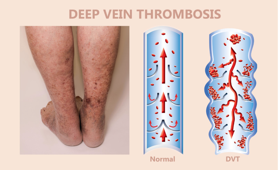
During the examination, the clinician initiated a sequence of measurements to compare the diameter of the affected calf with the healthy side. In the UK, this professional framework provides a stable foundation for the health journey by identifying that physical asymmetry is a primary physiological health factor in systemic surveillance. By utilised these integrated pathways, the healthcare system ensures every person profile is supported through evidence-based understanding of their vascular environment. This coordinated effort focuses on maintaining biological stability and detecting the mechanical triggers of fluid accumulation through regular specialist reviews to achieve long-term stability within the United Kingdom medical system.
The Wells Clinical Probability Score
The Wells score is a points-based clinical assessment tool utilised across the United Kingdom to determine the probability of an individual having a deep vein thrombosis. In the United Kingdom, healthcare professionals identify that categorising patients into “DVT likely” or “DVT unlikely” groups is essential for deciding the next sequence of diagnostic tests required to maintain biological homeostasis. NICE clinical guidelines indicate that the assessment of clinical probability using the Wells score is a central component of the DVT diagnostic pathway to maintain systemic stability.
| Wells Score Criteria | Points Awarded |
| Active Cancer | +1 Point |
| Pitting Oedema | +1 Point |
| Entire Leg Swollen | +1 Point |
| Calf Swelling >3cm | +1 Point |
| Previous History of DVT | +1 Point |
| Alternative Diagnosis Likely | -2 Points |
Identifying these clinical markers helps the multidisciplinary team provide a secure environment for health maintenance. In the UK, the focus is on providing a safe and accurate understanding of the individual functional capability across different life stages. This professional oversight is essential for building long-term health wellbeing through the systematic identification of circulatory triggers and the provision of specialist advice on achieving biological homeostasis during the diagnostic sequence within the national framework.
The Role of the D-dimer Blood Test
A D-dimer test is a specialised blood test utilised to identify fragments of a blood clot that has begun to break down within the circulatory system. In the United Kingdom, healthcare professionals identify that while a high D-dimer level does not prove a DVT exists, a negative result is highly effective at ruling out a clot in individuals with a low clinical probability. The GOV.UK health pages provide clinical profiles indicating that the monitoring of biological markers like D-dimer is a priority for ensuring integrated support through national safety programmes.
As the body interacts with a thrombus, the sequence of clotting and fibrinolysis leads to the release of these protein fragments. In the UK, the focus is on providing a stable foundation for the individual to move forward with self-understanding of their physical state. The NHS ensures that adults have a consistent point of contact for their health needs while they navigate the results of laboratory investigations. By utilised these integrated pathways, the healthcare system provides a secure environment for building long-term health wellbeing across the UK population. This integrated approach ensures that every person functional capability is respected and that clotting triggers are addressed through coordinated medical observation within the national framework.
Venous Ultrasound Imaging for Confirmation
The definitive diagnosis of deep vein thrombosis in the United Kingdom is usually achieved through a proximal leg vein ultrasound scan, which visualises the blood flow and the physical structure of the veins. In the United Kingdom, healthcare professionals focus on whether the veins can be compressed with the ultrasound probe, as a vein containing a solid clot will remain rigid and non-compressible.
Imaging features monitored in the UK include:
- Compressibility: Checking if the vein collapses under gentle pressure.
- Thrombus Visualisation: Identifying a solid mass within the vessel lumen.
- Doppler Flow: Assessing the speed and direction of blood movement.
- Vein Diameter: Measuring for abnormal expansion caused by a blockage.
- Respiratory Variation: Observing how blood flow changes with breathing.
- Clot Extent: Determining how far the thrombus extends up the leg or pelvis.
- Augmentation: Checking if manual calf squeezing increases blood flow.
In the UK, the focus is on providing a stable foundation for the individual to move forward with self-understanding. The NHS ensures that adults and children have a consistent point of contact for their health needs. By utilised these integrated pathways, the healthcare system provides a secure environment for building long-term health wellbeing across the UK population. These strategies aim to work with the individual’s biology to restore stability through the consistent clinical control of their environment and the systematic identification of the mechanical triggers of the circulatory system within the national framework.
Standard UK Protocols for Diagnostic Sequence
The United Kingdom healthcare system utilise a sequence of investigative protocols that vary depending on whether the initial Wells score suggests DVT is likely or unlikely. In the United Kingdom, healthcare professionals focus on ensuring that suspected thrombosis is investigated systematically to achieve biological homeostasis and provide an accurate physiological understanding of the vascular system state.
Investigative steps in the UK include:
- Initial Assessment: Performing a physical exam and calculating the Wells score.
- Blood Screening: Ordering a D-dimer test for those in the “unlikely” category.
- Urgent Imaging: Arranging a proximal leg vein ultrasound within 24 hours.
- Interim Treatment: Providing anticoagulant medication if a scan is delayed.
- Specialist Referral: Consulting with haematology or vascular teams for complex cases.
- Active Surveillance: Scheduled monitoring during clinical reviews to ensure stability.
- Systemic Screening: Checking for underlying causes if a clot is unprovoked.
In the UK, the focus is on providing a stable foundation for the individual to move forward with self-understanding. The NHS ensures that adults and children have a consistent point of contact for their health needs. By utilised these integrated pathways, the healthcare system provides a secure environment for building long-term health wellbeing across the UK population. These strategies aim to work with the individual’s biology to restore stability through the consistent clinical control of their circulatory health and the promotion of a sustainable lifestyle within a validated medical environment.
Conclusion
The diagnosis of deep vein thrombosis follows a sequence of clinical scoring, blood markers, and medical imaging designed to maintain vascular health within the established UK healthcare framework. The NHS and professional bodies provide a robust system of multidisciplinary assessments to help individuals achieve stability and resilience throughout their health journey. By focusing on both the consistent monitoring of circulatory health and the recognition of red flags, the system promotes the highest possible level of independence. Following a coordinated management plan with the help of medical experts ensures that unique adult needs are addressed holistically.
If you experience severe, sudden, or worsening symptoms, call 999 immediately.
What happens if my D-dimer test is positive?
In the UK, a positive D-dimer means you require a follow-up ultrasound scan to confirm whether a physical blood clot is present in the veins.
Is a leg ultrasound painful when checking for DVT?
In the UK, the scan is non-invasive and generally painless, though the clinician may apply firm pressure to check if your veins are compressible.
Can a GP diagnose DVT just by looking at my leg?
In the UK, while a GP can identify symptoms that suggest DVT, they must use the clinical pathway including scans or blood tests to confirm it.
Why do I need a repeat scan after one week?
In the UK, if your initial scan was negative but symptoms persist, a repeat scan may be needed to ensure a small clot has not grown or moved.
How long does it take to get the results of a DVT test?
In the UK, blood results are often available within hours, and the NHS aims to provide ultrasound scans for suspected DVT within 24 hours.
Will a normal X-ray show a blood clot in the leg?
No; in the UK, a standard X-ray only sees bones and some soft tissue, whereas a specialised ultrasound is required to see the blood vessels.
Who should I talk to if I suspect I have a blood clot?
The first point of contact in the United Kingdom is your GP or 111 for an urgent clinical assessment and to determine which tests are required.
Authority Snapshot (E-E-A-T)
This article provides medically factual health education regarding the diagnosis of DVT, strictly aligned with NHS and NICE clinical guidelines. The content is developed by a professional medical writing team and reviewed by Dr. Stefan Petrov, a UK-trained physician with experience in general surgery, emergency care, and intensive care. All information follows current UK public health protocols to ensure clinical accuracy and patient safety.



