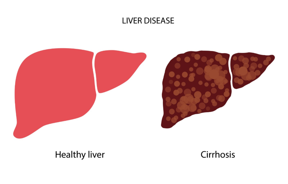
Monitoring for liver cancer in patients with cirrhosis is a structured process that occurs every six months to ensure the earliest possible detection of malignant changes. Cirrhosis involves extensive scarring of the liver, which significantly alters the cellular environment and increases the risk of hepatocellular carcinoma, the most common form of primary liver cancer. Because tumours can grow silently in a scarred liver without causing new physical symptoms, the UK healthcare system relies on a proactive surveillance pathway rather than waiting for a patient to feel unwell. This rigorous monitoring schedule is designed to identify abnormal growths when they are small and potentially curable. By adhering to a consistent cycle of clinical reviews, patients with advanced liver disease can significantly improve their long-term health outcomes and ensure that any complications are managed promptly.
What We’ll Discuss in This Article
- The biological rationale for the six-monthly surveillance interval.
- The primary diagnostic tools used during a liver cancer screening appointment.
- How multidisciplinary teams evaluate changes in liver nodules over time.
- The role of blood markers in supporting imaging results.
- Specific monitoring requirements for different causes of cirrhosis.
- The importance of consistent attendance in the NHS surveillance programme.
The Six-Monthly Surveillance Cycle
The standard interval for liver cancer surveillance in patients with cirrhosis is every six months because this timeframe aligns with the average doubling time of primary liver tumours. This frequent monitoring ensures that if a tumour begins to develop, it is likely to be identified while it is still less than two to three centimetres in size. The NHS states that if you have cirrhosis, you should have an ultrasound scan and sometimes a blood test every six months to check for liver cancer.
At this frequency, medical interventions such as surgical resection, ablation, or liver transplantation remain viable options. If the interval between scans is too long, there is a higher risk that a tumour could grow or spread beyond the liver before it is detected. For this reason, the six-monthly schedule is a non-negotiable standard of care for anyone in the United Kingdom with a confirmed diagnosis of liver cirrhosis.
Primary Tools for Cancer Detection
The core of the surveillance programme consists of an abdominal ultrasound scan, which provides a real-time visual assessment of the liver’s structure and any new growths. Ultrasound is a safe, non-invasive method that allows sonographers to identify nodules that may require further investigation. NICE clinical guidelines recommend six-monthly surveillance using abdominal ultrasound for people with cirrhosis to detect hepatocellular carcinoma at a treatable stage.
In addition to imaging, clinicians often perform a blood test to measure alpha-fetoprotein levels. This protein can act as a tumour marker, as its levels sometimes rise when liver cancer cells are present. While alpha-fetoprotein is not always elevated in early-stage cancer, it serves as a helpful supplementary tool when interpreted alongside the ultrasound images. If either the scan or the blood test shows an abnormality, the patient is usually referred for more detailed imaging, such as a CT or MRI scan, to confirm the nature of the growth.
Evaluating Nodules and Structural Changes
In a cirrhotic liver, it is common for the ultrasound to detect small lumps called regenerative nodules, which are usually harmless areas of healing tissue. The challenge for the clinical team is to distinguish these benign nodules from early-stage cancerous tumours. When a new nodule is found, its size and appearance are carefully documented and compared with previous scans to look for signs of growth or changes in blood supply.
Multi-disciplinary teams, consisting of radiologists, hepatologists, and surgeons, review these findings collectively. If a nodule is very small, the team may decide to monitor it more closely, perhaps increasing the frequency of scans temporarily. If the nodule meets certain criteria, such as a diameter over one centimetre or a specific blood flow pattern, the clinical pathway moves toward formal diagnosis and treatment planning. This systematic evaluation ensures that patients are not over-treated for harmless nodules while ensuring that malignancies are not missed.
Monitoring Variation by Cause of Cirrhosis
While the six-monthly interval is the standard for almost all patients with cirrhosis, the intensity of monitoring may be influenced by the underlying cause of the liver damage. Patients with cirrhosis caused by chronic viral hepatitis, particularly Hepatitis B, may require additional blood tests to monitor viral load alongside their cancer scans.
| Cause of Cirrhosis | Surveillance Goal | Frequency |
| Alcohol-Related | Early tumour detection and abstinence monitoring | Every 6 months |
| Fatty Liver (NAFLD) | Monitoring metabolic factors and tumour growth | Every 6 months |
| Viral Hepatitis (B/C) | Cancer detection and viral suppression checks | Every 6 months |
| Autoimmune Disease | Detecting nodules and managing inflammation | Every 6 months |
The GOV.UK health pages emphasise that regardless of the initial cause, the presence of cirrhosis itself is the primary trigger for entering the national liver cancer surveillance programme. Even if a patient has stopped drinking alcohol or has successfully cleared a Hepatitis C infection, the risk of cancer persists due to the existing scar tissue, making continued six-monthly monitoring essential for life.
The Importance of Consistent Attendance
The success of liver cancer prevention in the UK is heavily dependent on the patient’s commitment to attending every scheduled appointment. Missing even one scan can extend the gap between screenings to a year or more, which significantly increases the chance of a tumour reaching an advanced stage before detection.
The surveillance programme is designed to provide a continuous safety net. For many patients, the routine nature of these appointments provides reassurance that their condition is being managed with professional oversight. Healthcare providers work to ensure that these appointments are accessible, but the responsibility for maintaining the schedule ultimately rests on a partnership between the patient and their clinical team.
Conclusion
Cirrhosis must be monitored every six months using ultrasound scans and blood tests to provide the best chance of preventing advanced liver cancer. This specific interval is clinically necessary to catch tumours while they are small enough for curative treatment. Consistent participation in this surveillance programme is the most effective way to manage the long-term risks associated with advanced liver scarring. If you experience severe, sudden, or worsening symptoms, call 999 immediately.
What happens if I miss my six-monthly scan?
You should contact your clinic or GP to rebook immediately, as a longer gap between scans increases the risk of a tumour growing undetected.
Is an ultrasound enough to find all cancers?
Ultrasound is the primary screening tool, but if it shows anything unusual, you will be referred for a more detailed CT or MRI scan.
Why do I need a blood test as well as a scan?
The alpha-fetoprotein blood test provides an extra layer of information that can sometimes flag a problem before it is clearly visible on an ultrasound.
Will my monitoring ever stop if my liver stays stable?
No, because the scar tissue of cirrhosis is permanent, the risk of cancer remains, and surveillance is usually a lifelong commitment.
Is the monitoring the same for everyone with cirrhosis?
Yes, the six-monthly ultrasound is the standard UK protocol for all patients with a confirmed diagnosis of cirrhosis.
Can liver cancer be prevented entirely with these scans?
Surveillance does not prevent cancer from starting, but it is designed to find it at a stage where it can be treated or cured.
Do I need to fast before my surveillance ultrasound?
You are usually asked to fast for six hours before an abdominal ultrasound to ensure the liver and surrounding structures can be seen clearly.
Authority Snapshot (E-E-A-T)
This article provides medically factual health education regarding liver cancer surveillance, strictly aligned with NHS and NICE clinical guidelines. The content is developed by a professional medical writing team and reviewed by Dr. Rebecca Fernandez, a UK-trained physician with experience in internal medicine, surgery, and emergency care. All information follows current UK public health standards to ensure accuracy and patient safety.