




Additional tests used in polycythaemia vera investigations include advanced molecular screening for rare mutations, abdominal imaging to assess organ size, and bone marrow examinations to evaluate cellular architecture. In the United Kingdom, healthcare professionals utilise these specialist procedures to provide a comprehensive profile of the bone marrow factory when initial blood assays require further clarification. By utilised integrated NHS pathways, individuals receive a stable foundation for clinical review to maintain their systemic stability and functional independence within a validated medical environment focused on maintaining biological homeostasis and providing an accurate understanding of their haematological status through evidence-based specialist reviews.
What We’ll Discuss in This Article
- Advanced molecular testing for rare genetic variants.
- The role of abdominal ultrasound in assessing spleen health.
- Bone marrow aspiration and trephine biopsy procedures.
- Specialist assays for iron stores and uric acid levels.
- Oxygen saturation and lung function tests for differentiation.
- Accessing integrated UK support pathways for clinical assessments.
Advanced Molecular Testing for Rare Genetic Variants
If the common V617F mutation is not detected during initial screening, clinicians in the United Kingdom may perform advanced molecular assays to look for rarer mutations in exon 12 of the JAK2 gene. In the United Kingdom, clinical research highlights that approximately five per cent of patients with polycythaemia vera possess these less common genetic markers rather than the standard variant. The NHS states that while most people with polycythaemia vera have a change in the JAK2 gene, further specialist tests can help confirm the specific type.
These advanced genetic reviews ensure that individuals with atypical molecular profiles are correctly identified and integrated into the appropriate management pathway. In the UK, this professional framework provides a stable foundation for the health journey by identifying that molecular precision is a primary physiological health factor. By utilised these integrated pathways, the healthcare system ensures that every person’s profile is supported through evidence-based understanding of their genetic drivers. This coordinated effort prioritises the safety of the individual within a validated medical environment that focuses on maintaining biological stability and preventing the complications associated with uncontrolled cell production.
Abdominal Ultrasound and Organ Assessment
An abdominal ultrasound is frequently utilised to assess the size of the spleen and liver, as enlargement of these organs is a common physical marker of bone marrow overactivity. In the United Kingdom, specialists recognise that the spleen often grows larger (splenomegaly) because it attempts to filter the excessive volume of red blood cells or begins to produce blood cells itself. NICE clinical guidelines indicate that imaging of the abdomen is a useful supplementary investigation for patients with suspected myeloproliferative neoplasms to evaluate organ involvement.
| Feature | Healthy Spleen Size | Spleen Findings in PV |
| Typical Length | 10cm to 12cm. | Often exceeds 13cm (splenomegaly). |
| Physical Sensation | Not palpable under the ribs. | May be felt as fullness or pain. |
| Biological Role | Normal blood filtration. | Overworked from excessive cell mass. |
| Imaging Method | Ultrasound or CT scan. | Primarily ultrasound in the UK. |
In the UK, these biological markers are managed through integrated care plans that prioritise a person-centred approach. Identifying an enlarged spleen helps the multidisciplinary team provide a secure environment for health maintenance by monitoring for pressure on surrounding organs. This professional oversight is essential for providing a safe and accurate understanding of the individual’s functional capability. By utilised these clinical assessments, the healthcare system provides a secure environment for building long-term health wellbeing through the identification of haematological triggers.
Bone Marrow Aspiration and Trephine Biopsy
A bone marrow biopsy provides a direct assessment of the blood-producing environment by allowing a specialist haematopathologist to examine the marrow architecture and cellular density. In the United Kingdom, healthcare professionals utilised this procedure to identify “panmyelosis,” which is the characteristic increase in red cells, white cells, and megakaryocytes (platelet-producing cells). The GOV.UK health pages provide clinical profiles indicating that bone marrow examination is a priority for ensuring integrated support and accurate diagnosis of primary bone marrow disorders.
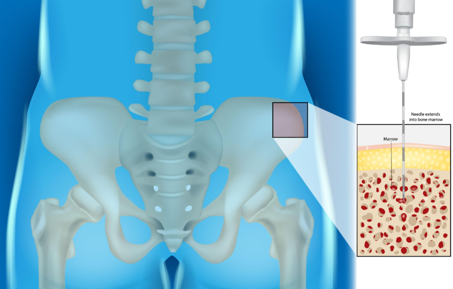
The procedure involves taking a liquid sample (aspiration) and a small solid core (trephine) from the pelvic bone. In the UK, the focus is on providing a stable foundation where the individual’s marrow health and fibre levels are reviewed regularly by a specialist team. Identifying these underlying drivers allows for more targeted help that addresses the actual biological cause of the erythrocytosis. By utilised these professional frameworks, the UK system provides a life-long framework of support that adapts to the person’s needs during different stages of adulthood.
Assays for Iron Stores and Uric Acid
Specialist blood tests to measure ferritin and uric acid levels are used to monitor the systemic impact of rapid cell turnover and frequent venesection. In the United Kingdom, healthcare professionals focus on these secondary markers because the bone marrow consumes large amounts of iron to produce red blood cells, which can lead to iron deficiency even when the red cell count is high.
Secondary markers monitored in the UK include:
- Serum Ferritin: Measuring the body’s iron stores, which are often depleted in PV.
- Uric Acid (Urate): Monitoring the byproduct of cell breakdown to prevent gout.
- Lactate Dehydrogenase (LDH): A marker of high cell turnover and marrow activity.
- Vitamin B12 Levels: Often elevated in primary bone marrow disorders.
- Liver Function Tests: Ensuring systemic health remains stable during treatment.
- Kidney Function Tests: Monitoring the organs responsible for hormone production.
In the UK, the focus is on providing a stable foundation for the individual to move forward with self-understanding of their metabolic health. The NHS ensures that adults have a consistent point of contact for their health needs while they navigate their lives. By utilised these integrated pathways, the healthcare system provides a secure environment for building long-term health wellbeing across the UK population. This integrated approach ensures that the person’s unique way of functioning is respected within their home and social environment.
Differentiation Through Oxygen and Lung Function
Tests such as pulse oximetry and formal lung function assessments are utilised to rule out secondary polycythaemia caused by chronic lung disease or low oxygen levels. In the United Kingdom, healthcare professionals must ensure that the high red blood cell count is not a physiological reaction to smoking or sleep apnoea before confirming a primary marrow condition.
Specific differentiation steps managed in the UK include:
- Pulse Oximetry: Measuring the percentage of oxygen saturation in the blood.
- Arterial Blood Gas: A more precise measure of oxygen and carbon dioxide.
- Spirometry: Assessing how much air a person can breathe out to check lung health.
- Sleep Studies: Identifying drops in oxygen at night that trigger cell production.
- Chest X-ray: Checking for heart or lung issues that might cause hypoxia.
- Echocardiogram: Ruling out heart defects that bypass oxygenation.
In the UK, the focus is on providing a stable foundation for the individual to move forward with self-understanding. The NHS ensures that adults and children have a consistent point of contact for their health needs. By utilised these integrated pathways, the healthcare system provides a secure environment for building long-term health wellbeing across the UK population. These strategies aim to work with the individual’s biology to restore a sense of purpose and stability.
Conclusion
The investigation of polycythaemia vera involves a detailed sequence of additional tests ranging from organ imaging to bone marrow reviews within the UK healthcare framework. The NHS and professional bodies provide a robust system of multidisciplinary assessments and reviews to help individuals achieve stability and resilience. By focusing on both the biological roots of the condition and the need for clinical oversight, the system promotes the highest possible level of independence. Following a coordinated management plan with the help of medical experts ensures that unique adult needs are addressed holistically.
Does everyone with PV need a bone marrow biopsy?
In most UK centres, a biopsy is recommended to confirm the diagnosis and provide a baseline for marrow health, although it may be omitted in specific circumstances.
Is an ultrasound painful?
No; an abdominal ultrasound is a painless procedure that uses sound waves to look at your internal organs.
What does it mean if my iron is low but my red cells are high?
In PV, your marrow uses up iron very quickly to make extra red cells, which often leads to low iron stores (ferritin).
Why do I need a lung function test?
The NHS needs to make sure your high blood count isn’t just your body’s way of coping with a lung problem or low oxygen.
How often will these additional tests be repeated?
Most imaging and biopsies are done at diagnosis, but blood markers like uric acid are checked regularly during your reviews.
Can a blood test alone tell the difference between PV types?
Specific genetic assays can identify whether you have the common JAK2 V617F mutation or a rarer variant.
Who should I talk to first if I am worried about an upcoming biopsy?
The first point of contact in the United Kingdom is usually your specialist haematology nurse or consultant at the hospital.
Authority Snapshot (E-E-A-T)
This article provides medically factual health education regarding additional tests for PV, strictly aligned with NHS and NICE clinical guidelines. The content is developed by a professional medical writing team and reviewed by Dr. Stefan Petrov, a UK-trained physician with experience in emergency care, surgery, and medical education. All information follows current UK public health protocols to ensure clinical accuracy and patient safety.



