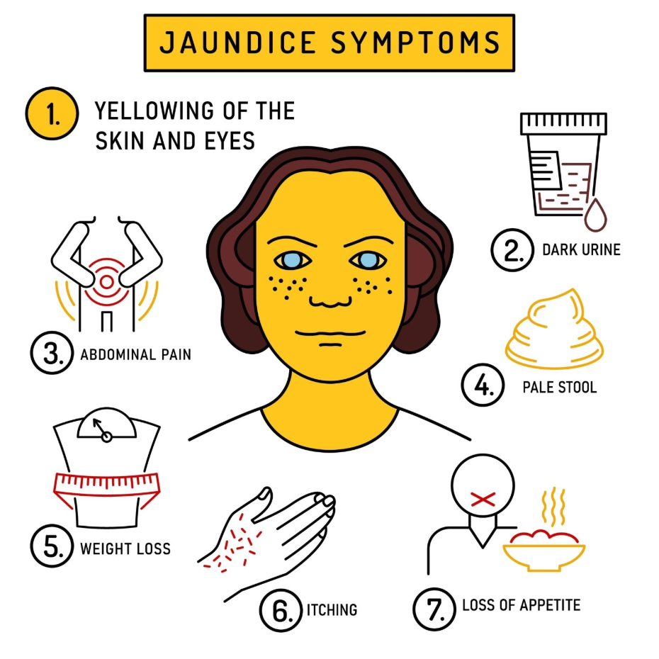
Jaundice is a medical sign characterised by a yellow discoloration of the skin and the whites of the eyes, a condition medically known as scleral icterus. This physical change occurs when there is an excess of a substance called bilirubin in the bloodstream, which then deposits into the body’s tissues. Under normal physiological conditions, the liver filters bilirubin from the blood and excretes it as a major component of bile. However, when the liver is unable to function correctly, or if there is a physical blockage in the drainage system, bilirubin levels rise and the pigment settles into tissues with high elastic content. Because the eyes are naturally white and contain specific proteins that bind to bilirubin, they are often the first part of the body where this accumulation becomes visible. In the United Kingdom, healthcare professionals use the appearance of the eyes as a primary indicator for assessing liver, gallbladder, and haematological health. Understanding the biological mechanism behind this yellowing is essential for identifying when the body’s waste management systems are under stress and require clinical investigation.
What We’ll Discuss in This Article
- The biological origin of bilirubin from the recycling of red blood cells.
- The liver’s role in filtering, conjugating, and excreting waste pigments.
- Why the sclera of the eye is particularly susceptible to yellowing.
- The clinical significance of yellow eyes as an early diagnostic sign.
- How different types of jaundice affect the body’s tissues and waste.
- The process of clinical investigation for new yellowing of the eyes.
The Role of Bilirubin in Tissue Discoloration
The whites of the eyes turn yellow because bilirubin, a yellow pigment created during the breakdown of red blood cells, has a high chemical affinity for the elastin found in the eye’s outer layer. The NHS states that jaundice is caused by the build-up of bilirubin in the blood, which can happen if the liver is damaged or there is a problem with the bile ducts.
Bilirubin is a natural byproduct of the body’s process for recycling aged red blood cells, which typically have a lifespan of about 120 days. When these cells are broken down in the spleen and liver, haemoglobin is converted into unconjugated bilirubin. This form of the pigment is fat-soluble and must be transported to the liver, where it is made water-soluble through a process called conjugation. Once conjugated, it can be safely excreted through the bile ducts into the intestines. When this metabolic pathway is interrupted, the concentration of bilirubin in the blood rises significantly. Because bilirubin binds easily to elastin, a protein abundant in the sclera, the eyes change colour as the pigment accumulates.
Why the Eyes Show Jaundice First
The eyes are often the first part of the body to show signs of jaundice because the sclera is naturally white, providing a stark contrast that makes the yellow pigment visible even at relatively low concentrations. In the early stages of a rise in bilirubin, the yellowing may not be noticeable on the skin, especially in individuals with darker skin tones or significant sun exposure. However, the lack of natural pigmentation in the whites of the eyes allows the yellow hue to be detected by both the patient and the clinician during a physical examination.
This early visibility makes the eyes a vital diagnostic tool in the United Kingdom health system. Scleral icterus often serves as the initial trigger for a medical review before more systemic symptoms, such as dark urine or abdominal discomfort, become prominent. Because the eyes are highly vascularised, meaning they have a rich supply of small blood vessels, the bilirubin pigment reaches these tissues rapidly once it has accumulated in the circulatory system. This allows for the early detection of liver or biliary stress, often before the liver’s functional capacity has significantly declined.
Anatomy of the Sclera and Pigment Binding
The sclera is the opaque, fibrous, outer protective layer of the human eye, and its unique protein composition is what causes it to trap bilirubin so effectively. It is composed primarily of collagen and elastin fibres. Elastin is a highly flexible protein that allows tissues to resume their shape after stretching, and it has a specific chemical attraction to bilirubin molecules.
When bilirubin levels in the blood exceed the normal range, the pigment leaks out of the capillaries and binds firmly to these elastin fibres. Unlike the skin, which has multiple layers of epidermis and varying levels of melanin, the sclera is covered only by a thin, transparent layer called the conjunctiva. This allows the yellowed elastin fibres underneath to be seen clearly. The combination of high elastin content and a transparent covering is the reason why the eyes are a more sensitive indicator of jaundice than almost any other part of the human anatomy.
Comparing Sclera and Skin Discoloration
While both skin and eye discoloration stem from the same underlying buildup of bilirubin, the intensity and timing of these changes can vary based on the individual’s physiology and the specific cause of the jaundice. Clinical professionals use these differences to assess the severity of the condition.
| Feature | Sclera (Whites of Eyes) | Skin |
| Tissue Affinity | High affinity due to elastin | Moderate affinity |
| Ease of Detection | High (white background) | Variable (affected by skin tone) |
| Timing of Appearance | Usually the first sign | Often develops later |
| Clinical Term | Scleral Icterus | Jaundice |
NICE clinical guidelines for jaundice in adults emphasise that a thorough examination of the sclera is essential for identifying subclinical jaundice that might not yet be visible on the skin surface. This focused examination ensures that metabolic issues are identified as early as possible, allowing for timely intervention.
Categories of Jaundice and Their Impact
Yellowing of the eyes can be triggered by three main categories of medical issues, classified by where the disruption occurs in the bilirubin cycle. These are known as pre-hepatic, intra-hepatic, and post-hepatic jaundice. Each category affects the body differently, though the yellowing of the eyes remains a constant feature.
Pre-hepatic jaundice occurs when red blood cells are destroyed too quickly, often due to blood disorders like anaemia. Intra-hepatic jaundice is caused by damage directly to the liver tissue, such as from viral hepatitis or alcohol-related liver disease, which prevents the organ from processing bilirubin. Post-hepatic jaundice, also known as obstructive jaundice, occurs when a physical blockage like a gallstone prevents the bile from draining into the intestines. In the United Kingdom, obstructive jaundice is often characterised by more intense yellowing of the eyes accompanied by pale stools and dark urine. According to the GOV.UK health pages, jaundice requires clinical assessment to rule out serious obstructions or advanced liver failure.
Clinical Investigation of Yellowing Eyes
When a patient presents with yellowing of the eyes in the UK, clinicians follow a structured diagnostic pathway to determine the cause. This begins with a physical examination and a review of other symptoms, such as itching, fatigue, or changes in waste products. Blood tests, known as liver function tests, are essential for measuring the levels of different types of bilirubin and liver enzymes. These tests help determine if the problem is rooted in the blood, the liver cells, or the bile ducts.
If an obstruction is suspected, imaging such as an ultrasound or CT scan is used to look for gallstones or other blockages in the biliary system. Because the yellowing of the eyes is a systemic sign, the investigation often involves a multidisciplinary team to ensure all aspects of the patient’s health are considered. Once the underlying cause is identified and treated, the bilirubin levels in the blood will begin to fall, and the yellow pigment will gradually clear from the eyes and skin.
Conclusion
The whites of the eyes turn yellow during jaundice because bilirubin pigment binds to the elastin proteins in the sclera. This symptom is frequently the first visible indicator of a liver, blood, or gallbladder issue, as the white background of the eye makes the discoloration easy to detect even at low concentrations. In the UK, healthcare professionals use the appearance of the eyes as a primary clinical marker to initiate investigations into the body’s filtration and excretion systems. Identifying this change early is essential for determining the underlying cause and supporting long-term health. If you experience severe, sudden, or worsening symptoms, call 999 immediately.
Is it possible for only one eye to turn yellow in jaundice?
No, jaundice is a systemic condition caused by a buildup of bilirubin in the blood, so it will always affect both eyes equally.
Can eye drops clear the yellowing caused by jaundice?
No, eye drops cannot remove the yellow colour because the pigment is deposited deep within the eye tissue from the bloodstream.
How high must bilirubin levels be to see it in the eyes?
Bilirubin levels usually need to be at least twice the normal limit before the yellowing becomes visible in the sclera.
Does yellowing of the eyes always mean liver failure?
Not necessarily, as yellowing can also be caused by temporary issues like gallstones or certain blood conditions.
Why do some people’s eyes look yellow without having jaundice?
Some individuals have harmless fatty deposits on the eye surface, called pinguecula, which can appear yellow but are not related to bilirubin.
Does the yellowing go away after the cause is treated?
Yes, once the underlying cause is resolved and bilirubin levels in the blood return to normal, the pigment will gradually be cleared from the eyes.
Can tiredness or lack of sleep turn the eyes yellow?
Tiredness may make the eyes look bloodshot or dull, but it will not cause the distinct yellowing of the sclera seen in jaundice.
Authority Snapshot (E-E-A-T)
This article provides medically factual health education regarding why jaundice affects the eyes, strictly aligned with NHS and NICE clinical guidelines. The content is developed by a professional medical writing team and reviewed by Dr. Stefan Petrov, a UK-trained physician with experience in general medicine, surgery, and emergency care. All information follows current UK public health protocols to ensure clinical accuracy and patient safety.