




Diverticulitis requires follow-up investigations, such as a scan or colonoscopy, once the acute inflammatory phase has resolved to ensure that no underlying colon polyps were hidden by the swelling of the bowel wall. While diverticulitis and colon polyps are distinct conditions involving different biological processes, they frequently occur in the same age group and can present with overlapping symptoms like rectal bleeding. In the United Kingdom, healthcare professionals follow a structured pathway where the immediate focus is on resolving the infection in the diverticular pouches before performing a thorough visual or radiological inspection of the colon lining. This approach is essential because the inflammation during a flare-up can make the bowel wall fragile and difficult to examine safely. Once the tissue has healed, typically after several weeks, a follow-up investigation allows clinicians to differentiate between the structural changes of diverticular disease and the cellular overgrowths of polyps. By ensuring the colon is clear of polyps after a diverticulitis episode, the UK healthcare system provides comprehensive preventative care and reduces the risk of future gastrointestinal complications. Understanding the timing and necessity of these follow-up checks is a vital part of managing long-term bowel stability.
What We’ll Discuss in This Article
- Why immediate investigations are postponed during an acute flare-up.
- The typical timeframe for follow-up scans or colonoscopies after infection.
- How inflammation can obscure the detection of colon polyps.
- The role of CT colonography as an alternative to invasive procedures.
- Differentiating between diverticular symptoms and signs of polyps.
- UK clinical protocols for ensuring a clear colon after diverticulitis.
Investigating the Colon After an Acute Flare-up
Individuals require a follow-up investigation after an episode of diverticulitis to confirm that their symptoms were purely inflammatory and to check for polyps that may have been present simultaneously. Diverticulitis occurs when small pouches in the colon wall, known as diverticula, become infected or inflamed. The NHS states that diverticulitis happens when the small pouches in the lining of the large intestine become inflamed or infected, and it is more common as people get older.
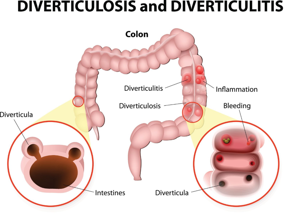
Because many people in the UK over the age of 50 have both diverticula and polyps, it is common for a clinician to find both during a routine check. However, if a patient presents with sudden abdominal pain and fever, the immediate clinical priority is to treat the infection. A follow-up check is necessary because symptoms like minor rectal bleeding, which are often attributed to diverticular disease, could also be caused by a “silent” polyp. By performing a scan or colonoscopy after the inflammation has cleared, healthcare providers can ensure that every structural issue within the large intestine is identified and managed appropriately according to its specific risk profile.
The Importance of Waiting for Tissue Healing
Clinical investigations to rule out polyps are typically delayed for six to eight weeks after a diverticulitis flare-up to allow the bowel wall to heal and to reduce the risk of procedural complications. During an active infection, the lining of the colon becomes swollen and the pouches can become very thin and fragile. NICE clinical guidelines for diverticular disease management indicate that invasive bowel tests like a colonoscopy should generally be avoided during an acute flare-up due to the higher risk of causing a perforation.
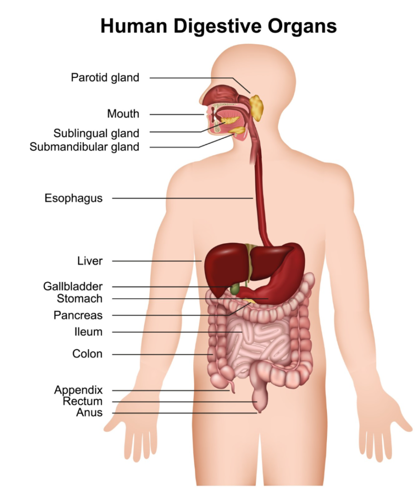
If a colonoscopy were performed while the tissue was still inflamed, the pressure from the air used to inflate the bowel could cause a weakened pouch to burst. Furthermore, the swelling (oedema) makes it difficult for the clinician to get a clear view of the mucosa, meaning small polyps could easily be missed. Waiting for the inflammatory response to subside ensures that the bowel is stable enough for a safe and thorough examination. This structured delay is a standard safety protocol in the United Kingdom, designed to provide the most accurate diagnostic results once the immediate crisis has been resolved with rest or antibiotics.
How Inflammation Obscures Polyp Detection
Inflammation in the colon wall can physically mask the presence of polyps by creating folds or swelling that hide small tissue growths from the view of a camera or a scanner. When a diverticulum is infected, the surrounding area of the bowel wall thickens as part of the body’s immune response. This thickening can make the inner surface of the colon appear irregular, which might hide a flat or small polyp.
| Feature | Diverticulitis Flare-up | Healthy Colon Lining |
| Wall Thickness | Swollen and thickened. | Thin and flexible. |
| Visual Clarity | Reddened and obscured by mucus. | Pale pink and clear. |
| Risk of Procedure | High risk of tissue tearing. | Standard low-risk environment. |
| Polyp Visibility | Poor; growths can be hidden in folds. | High; protrusions are easily seen. |
Because the goal of a follow-up check is to ensure a completely “clear” colon, performing the scan too early could lead to a false sense of security if a growth is missed. Once the swelling has gone down, the bowel wall returns to its normal thickness, allowing the clinician to perform a much more precise visual inspection. In the UK, this ensures that the preventative goals of bowel screening identifying and removing polyps before they can change are met effectively.
The Role of CT Colonography as an Alternative
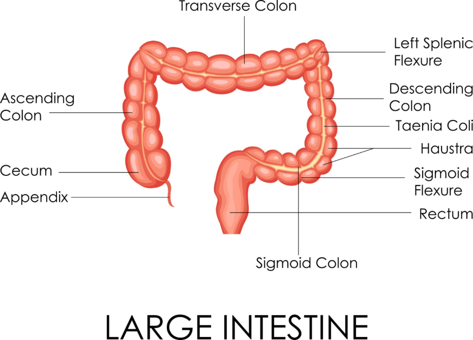
CT colonography, often called a virtual colonoscopy, is a scan that may be used to rule out polyps in patients who have recovered from diverticulitis but are not suitable for a traditional colonoscopy. This test uses a CT scanner to create detailed three-dimensional images of the colon and is particularly useful if the bowel remains slightly narrowed or “loopy” following a severe episode of diverticulitis.
NICE clinical guidelines for colorectal cancer indicate that CT colonography is an effective diagnostic tool for detecting polyps and is an appropriate alternative for patients where a full colonoscopy is not clinically possible. While this scan is non-invasive, it still requires the patient to follow a bowel-cleansing preparation to ensure the images are clear. It is excellent for mapping the structural health of the colon wall and identifying larger polyps. If a significant growth is detected on the scan, the patient will then be referred for a targeted colonoscopy to have the tissue removed. This integrated use of imaging technology ensures that all patients, regardless of the severity of their diverticular disease, have a pathway to a clear colon.
Differentiating Symptoms for Targeted Investigation
A follow-up scan is especially important if a patient continues to experience symptoms like rectal bleeding or altered bowel habits after their diverticulitis infection has supposedly cleared. While diverticulitis causes acute pain and fever, polyps are often “silent” or cause only minor, intermittent bleeding. If these symptoms persist once the inflammatory markers in the blood have returned to normal, it is a clinical sign that something other than diverticula may be affecting the bowel.
In the United Kingdom, any unexplained change in bowel habits lasting three weeks or more is a trigger for a more detailed review. By ruling out polyps through a scan or colonoscopy, clinicians can confidently attribute any remaining minor symptoms to chronic diverticular disease and focus on dietary management. This prevents a scenario where a patient dismisses a sign of a new growth as just another flare-up of their known diverticular pouches. Maintaining this diagnostic distinction is a cornerstone of UK gastrointestinal health, ensuring that the appropriate treatment is provided for both infectious and cellular bowel issues.
UK Clinical Pathways and Patient Safety
The management of diverticulitis in the United Kingdom follows a logical progression that prioritises immediate safety and long-term preventative health through staged investigations. Once a patient has recovered from an acute episode, they will typically have a review with their GP or a specialist to discuss the timing of their follow-up check. The GOV.UK health pages indicate that a structured follow-up after an acute bowel event is essential for ensuring that no other health concerns, such as polyps, are overlooked.
This integrated approach ensures that the patient is not just treated for the “loud” symptoms of an infection but is also screened for the “silent” risks of cellular overgrowths. The results of the follow-up investigation will determine the future monitoring schedule. If polyps are found and removed, the patient enters a surveillance programme; if only diverticula are present, the focus remains on high-fibre dietary management to prevent future flare-ups. By adhering to these national clinical protocols, the UK healthcare system provides a comprehensive safety net that protects the functional and structural health of the large intestine throughout the patient’s life.
Conclusion
Diverticulitis requires a follow-up scan or colonoscopy several weeks after an acute flare-up to ensure that the colon is clear of polyps that may have been obscured by inflammation. While the immediate focus is on treating the infection, the staged investigation is vital for long-term preventative health and for differentiating between different types of bowel structural changes. In the UK, these protocols prioritise patient safety by waiting for the bowel wall to heal before performing detailed checks. Maintaining a high-fibre diet and participating in all scheduled follow-ups are the most effective ways to manage the risks associated with both conditions. If you experience severe, sudden, or worsening symptoms, call 999 immediately.
Why can’t I have a scan immediately to check for polyps?
During a flare-up, inflammation and swelling make it difficult to see small polyps clearly, and the bowel wall is too fragile for invasive checks.
Is a CT scan enough to rule out polyps?
A CT colonography is very good at finding larger polyps, but a colonoscopy is the only way to see the lining directly and remove any growths found.
How long should I wait after my pain goes away before having a check-up?
UK clinical guidelines usually recommend waiting six to eight weeks after your symptoms have completely resolved before undergoing a follow-up colonoscopy.
Will my diverticula disappear after the infection is treated?
No, diverticula are permanent structural changes to your bowel wall, but the inflammation (diverticulitis) will resolve with appropriate care.
Can a home screening kit replace the follow-up scan?
No; the home kit only looks for hidden blood and cannot provide the detailed visual check needed to ensure your colon is clear after an infection.
What if my follow-up colonoscopy is normal?
A normal result means no polyps were found; you will then focus on dietary management to prevent your diverticula from flaring up again.
Does everyone with diverticulitis need a follow-up scan?
In the UK, a follow-up investigation is typically recommended for most people after their first episode of diverticulitis to confirm the diagnosis and check for other issues.
Authority Snapshot (E-E-A-T)
This article provides medically factual health education regarding the timing of scans for polyps following diverticulitis, strictly aligned with NHS and NICE clinical guidelines. The content is developed by a professional medical writing team and reviewed by Dr. Stefan Petrov, a UK-trained physician with extensive experience in emergency care, surgery, and general medicine. All information follows current UK public health protocols to ensure clinical accuracy and patient safety.



