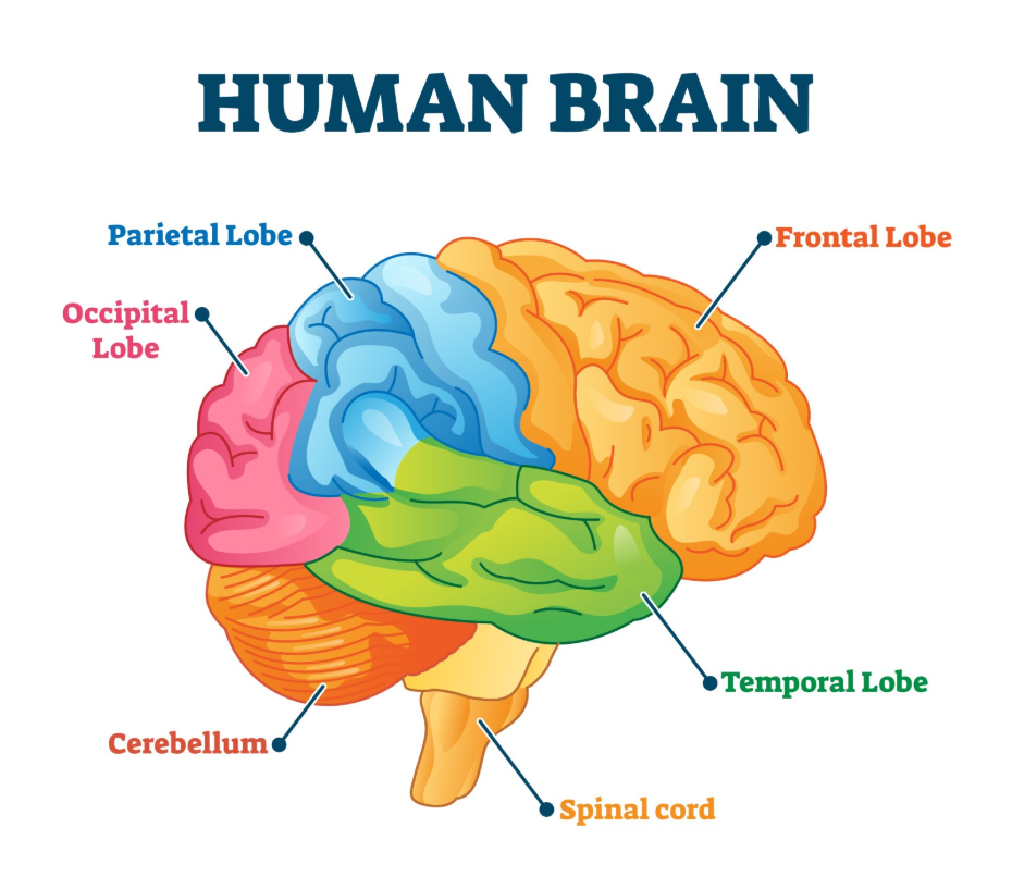
Early detection of a brain tumour can influence the clinical outcome by allowing for medical intervention before the growth causes significant neurological damage or increased intracranial pressure. In the United Kingdom, healthcare professionals utilise structured referral pathways to ensure that patients with suspicious symptoms receive prompt neuroimaging and specialist assessment. While the biological type and grade of the tumour remain primary factors in the long-term prognosis, identifying a mass at an earlier stage may provide more options for surgical removal or targeted therapies. The NHS follows evidence-based protocols established by NICE to prioritise urgent cases and streamline the diagnostic journey. Understanding the clinical significance of early identification helps patients and families navigate the healthcare system with a focus on timely and accurate assessment. This article explores the impact of timing on treatment efficacy, the role of red-flag symptoms, and the integrated safety nets provided within the UK healthcare framework to support patients throughout the diagnostic process.
What We’ll Discuss in This Article
- The relationship between tumour size at detection and surgical feasibility.
- How early identification helps preserve neurological and functional health.
- The role of “red-flag” symptoms in triggering urgent NHS referrals.
- Understanding the 28-day faster diagnosis standard in the United Kingdom.
- The impact of early intervention on the management of low-grade tumours.
- How multidisciplinary teams coordinate rapid assessment and care planning.
Impact of Detection Timing on Surgical Options
Early detection can improve the likelihood of a successful surgical outcome because tumours identified when they are smaller and more contained are often easier for neurosurgeons to remove safely. When a tumour is found before it has heavily infiltrated surrounding healthy brain tissue or intertwined with critical blood vessels, the surgeon has a better opportunity to achieve a maximal safe resection. The NHS states that a brain tumour diagnosis typically begins with a GP who will check your vision, hearing, and coordination before making a referral.
In the United Kingdom, the primary goal of surgery is to remove as much of the abnormal growth as possible while protecting vital brain functions such as speech and movement. If a tumour is detected late, it may have grown to a size where its removal carries a higher risk of causing permanent disability. Early identification allows the multidisciplinary team to plan the surgical approach with greater precision, potentially using less invasive techniques. By reducing the “tumour burden” at an earlier stage, subsequent treatments like radiotherapy or chemotherapy may also be more effective in managing any remaining microscopic cells.
Preserving Neurological and Functional Health
Identifying a brain tumour early is vital for preserving a patient’s neurological function, as it allows for management to begin before the mass causes irreversible damage to sensitive neural pathways. As a tumour grows, it exerts pressure on the surrounding brain tissue, which can lead to a range of deficits including vision loss, speech difficulties, or physical weakness. NICE clinical guidelines for brain tumours indicate that healthcare professionals should consider an urgent referral for suspected brain cancer if a patient presents with a progressive neurological deficit or a new-onset seizure.
Early intervention aims to “decompress” the brain and alleviate this pressure before the damage becomes permanent. In the United Kingdom, specialist neuro-rehabilitation services are integrated into the care pathway to support patients who do experience functional changes. However, starting treatment while the patient still has a high level of physical and cognitive health generally leads to a smoother recovery and a better overall quality of life. By focusing on the preservation of function, the NHS prioritises the patient’s long-term independence. Early detection provides the best window of opportunity for clinicians to implement strategies that protect the delicate balance of the central nervous system.
The Role of Red-Flag Symptoms in the UK
In the United Kingdom, early detection is facilitated by the identification of “red-flag” symptoms which signal to GPs that a patient requires urgent specialist investigation rather than routine monitoring. These indicators help move patients past common medical mimics, such as standard tension headaches or stress-related fatigue, and into the neuro-oncology pathway.
Key red-flag indicators monitored by UK clinicians include:
- Persistent Morning Headaches: Especially if they are accompanied by nausea or vomiting.
- New-onset Seizures: Any seizure activity in an adult with no previous history of epilepsy.
- Progressive Weakness: A gradual loss of strength or sensation on one side of the body.
- Unexplained Vision Changes: Such as double vision or a loss of peripheral sight.
- Significant Personality Shifts: Rapid changes in mood, behaviour, or cognitive ability.
By recognising these specific signs, healthcare providers can initiate an urgent referral under the faster diagnosis standards. This systematic approach ensures that high-risk cases are not overlooked. Patients are encouraged to provide a clear timeline of their symptoms to their GP, as the “progressive” nature of these signs is often a critical factor in determining the urgency of the referral. Identifying these markers early is a cornerstone of the UK strategy to improve neurological outcomes.
Understanding the 28-Day Faster Diagnosis Standard
The United Kingdom has implemented the 28-day faster diagnosis standard to ensure that patients with suspected brain tumours receive a definitive diagnosis or a confirmation that cancer is not present within four weeks of an urgent referral. This standard is designed to reduce the anxiety of waiting for results and to ensure that treatment can begin as soon as possible if a tumour is identified. The GOV.UK health pages provide clinical profiles indicating that meeting diagnosis targets is a key priority for improving cancer survival and patient experience in the UK.
During this 28-day window, the NHS coordinates:
- Urgent Imaging: Providing rapid access to CT or MRI scans.
- Specialist Review: Ensuring the results are evaluated by a neuroradiologist and a neurosurgeon.
- Biopsy (if required): Performing a tissue sample to confirm the tumour type and grade.
- MDT Discussion: Reviewing the case within a multidisciplinary team to finalise the management plan.
This integrated framework removes many of the historical barriers to early detection. By centralising the diagnostic process, the UK healthcare system ensures that patients move through the necessary tests efficiently. Early detection through this streamlined pathway means that the clinical team can act while the tumour is at its most manageable stage, providing the best possible foundation for the subsequent phases of care.
Management of Low-Grade tumours and Transformation
Early detection is particularly significant for low-grade brain tumours, as it allows for long-term monitoring and early intervention to manage the risk of “malignant transformation.” Some low-grade tumours grow very slowly and may be managed through a “watch and wait” approach, but they have the potential to become more aggressive over several years.
| Aspect of Early Detection | Benefit for Low-Grade tumours | Clinical Rationale |
| Active Surveillance | Establishes a baseline early. | Allows clinicians to track even subtle growth. |
| Surgical Timing | removal before infiltration. | Prevents the tumour from spreading into vital tissue. |
| Seizure Control | Prompt medication. | Improves quality of life and safety early on. |
| Transformation Check | Early catch of grade changes. | Ensures treatment is stepped up as soon as needed. |
In the United Kingdom, detecting a low-grade tumour early means that the multidisciplinary team can establish a regular scanning schedule. This consistent surveillance ensures that if the tumour shows signs of changing into a higher grade, the medical team can intervene with radiotherapy or surgery immediately. This proactive management is essential for extending the period of stable health for the patient. By catching these changes at the earliest opportunity, the NHS aims to delay or prevent the onset of more serious neurological complications associated with high-grade disease.
Collaborative Care and Diagnostic Accuracy
Achieving an early and accurate diagnosis in the United Kingdom involves a collaborative effort between primary care, radiology, and the neuro-oncology multidisciplinary team. This group of experts ensures that every scan and test result is interpreted with a high degree of specialist knowledge, reducing the risk of a mass being misidentified or overlooked.
The collaborative diagnostic process includes:
- Neuroradiology Expertise: Specialist doctors interpreting complex brain images.
- Pathology Analysis: Examining tissue samples to identify specific molecular markers.
- Surgical Assessment: Evaluating if an early operation is the safest management route.
- Neurological Monitoring: Assessing the impact of the tumour on the patient’s daily function.
This team-based approach provides a vital safety net for patients. In the UK, if an initial scan is inconclusive, the team may recommend a follow-up scan within a short timeframe rather than waiting for symptoms to worsen. This vigilance is a key part of the early detection strategy within the NHS. By bringing together multiple specialities, the healthcare system ensures that the diagnosis is not just fast, but also precise, providing a clear and evidence-based plan for the patient’s future care.
Conclusion
Early detection can improve the outcome of a brain tumour by allowing for surgical removal while the mass is smaller and preserving vital neurological function before damage occurs. In the UK, the NHS uses red-flag symptoms and the 28-day faster diagnosis standard to ensure that patients are assessed and diagnosed promptly. While the biological grade of the tumour is a major factor in the prognosis, early identification provides a wider range of management options and better functional preservation. Multidisciplinary teams coordinate this rapid assessment to ensure diagnostic accuracy and timely care. Consistent monitoring and follow-up are essential parts of the clinical journey in the United Kingdom. If you experience severe, sudden, or worsening symptoms, call 999 immediately.
Does a headache always mean I need an urgent brain scan?
No; most headaches are not related to tumours, but UK GPs look for “red-flag” features like morning pain or associated nausea before referring for a scan.
Can an eye test detect a brain tumour early?
Yes; an optician can sometimes see swelling at the back of the eye (papilledema) which indicates pressure in the brain and requires an urgent referral.
Will early detection guarantee a cure for a brain tumour?
Early detection provides more options and better function, but the possibility of a cure depends on the specific tumour type and its response to treatment.
How long does it take to get scan results in the UK?
Under the 28-day faster diagnosis standard, the NHS aims to provide you with a diagnosis or an update within four weeks of an urgent referral.
Is a CT scan as good as an MRI for early detection?
A CT scan is excellent for identifying large masses or bleeding, but an MRI provides more detailed images that are often needed for a precise diagnosis.
Why do some tumours take longer to detect than others?
Slow-growing tumours may cause very subtle symptoms that only become apparent over several months, making them harder to identify initially.
What should I do if my GP says my symptoms are just stress?
In the UK, if your symptoms persist or worsen, you should return to your GP for a review, as the “progressive” nature of symptoms is a key diagnostic indicator.
Authority Snapshot (E-E-A-T)
This article provides medically factual health education regarding the impact of early detection on brain tumour outcomes, strictly aligned with NHS and NICE clinical guidelines. The content is developed by a professional medical writing team and reviewed by Dr. Rebecca Fernandez, a UK-trained physician with experience in general surgery, cardiology, emergency medicine, and psychiatry. All information follows current UK public health protocols to ensure clinical accuracy and patient safety.
