Imaging scans are essential clinical tools used to identify and monitor various liver conditions, offering a non-invasive way to visualise the organ’s structure, fat content, and the presence of abnormal growths. While technologies such as ultrasound, computed tomography, and magnetic resonance imaging provide high levels of detail, their accuracy can vary depending on the specific stage of the disease and the characteristics of the patient. In the United Kingdom, the NHS uses a structured approach where different types of scans are selected based on their specific strengths, such as measuring tissue stiffness or identifying the blood supply to a suspected tumour. These scans allow healthcare professionals to track the progression from simple fat accumulation to advanced scarring without the immediate need for more invasive procedures. Understanding the capabilities and limitations of each imaging modality is vital for patients as they navigate the diagnostic pathway and participate in long term surveillance programmes.
What We’ll Discuss in This Article
- The accuracy of ultrasound in detecting fat accumulation.
- How transient elastography measures liver stiffness and scarring.
- The role of CT and MRI scans in identifying liver cancer.
- Limitations of imaging in detecting early-stage liver damage.
- Comparison of different scan types for specific liver conditions.
- How UK clinical guidelines determine the choice of imaging.
Ultrasound Accuracy in Detecting Fatty Liver
Ultrasound is the primary tool used in the UK for the initial detection of fatty liver disease because it is highly accessible and effective at identifying significant fat accumulation. During the scan, fat appears as a bright or echogenic area, allowing the sonographer to see a clear contrast between fatty tissue and healthy liver tissue. The NHS states that an ultrasound scan can be used to show if you have a fatty liver, although it cannot always tell how much scarring is present.
The accuracy of a standard ultrasound is highest when the fat content of the liver exceeds twenty to thirty percent. In cases where the fat accumulation is very mild, the scan may appear normal, which is a known limitation of this technology. Additionally, ultrasound is less accurate at distinguishing between simple fat and active inflammation. Despite these limitations, it remains the standard first-line investigation in the UK for patients with metabolic risk factors, providing a reliable starting point for further clinical evaluation.
Measuring Scarring with Transient Elastography
Transient elastography, commonly referred to by the brand name FibroScan, is highly accurate at measuring liver stiffness, which is a direct indicator of cirrhosis and advanced fibrosis. This specialised ultrasound technology measures the speed of a shear wave as it travels through the liver; the faster the wave travels, the stiffer the liver tissue, suggesting more extensive scarring. NICE clinical guidelines recommend the use of transient elastography to diagnose cirrhosis in adults with chronic liver disease because of its high diagnostic accuracy compared to traditional imaging.
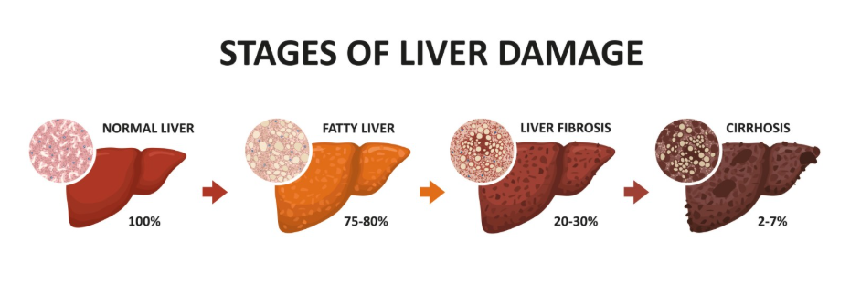
The FibroScan is particularly valued for its ability to identify “silent” cirrhosis in patients who may have normal blood tests and no physical symptoms. While it is very accurate for detecting advanced stages of scarring, its precision can sometimes be affected by factors such as acute liver inflammation or significant obesity. In the UK, it has largely replaced the need for liver biopsies in routine clinical practice, providing a safe and repeatable way to monitor how liver stiffness changes over time.
Detection and Characterisation of Liver Cancer
For the detection of liver cancer, imaging accuracy depends on the ability to identify small tumours and distinguish them from harmless regenerative nodules. Abdominal ultrasound is used for six-monthly surveillance in high-risk patients, but if an abnormality is found, more detailed scans such as Computed Tomography or Magnetic Resonance Imaging are required for a definitive conclusion. These advanced scans use contrast dyes to highlight the unique blood flow patterns that are characteristic of liver cancer.
| Scan Type | Accuracy for Cancer | Key Advantage |
| Ultrasound | Moderate (Screening) | Safe, no radiation, used for regular checks |
| CT Scan | High (Diagnostic) | Fast, excellent for identifying tumour size |
| MRI Scan | Very High (Diagnostic) | Best for soft tissue detail and small nodules |
An MRI is often considered the most accurate imaging modality for detecting very small liver tumours, especially in a liver that is already heavily scarred by cirrhosis. CT scans are also highly accurate and are frequently used to see if the cancer has spread to nearby blood vessels or other organs. The choice between these scans in the UK is made by a multidisciplinary team to ensure the highest degree of diagnostic certainty.
Limitations and Factors Affecting Scan Accuracy
While modern imaging is sophisticated, several factors can influence the accuracy of the results, sometimes leading to a need for repeat scans or different modalities. Body composition is a significant factor, as excess abdominal fat can sometimes make it difficult for ultrasound waves to reach the liver tissue clearly. Similarly, the presence of fluid in the abdomen can occasionally obscure the view of the liver surface during a standard scan.
Early-stage liver damage is also difficult to detect accurately with imaging. Simple inflammation (non-alcoholic steatohepatitis) and early-stage fibrosis may not cause enough structural change to be visible on an ultrasound or CT scan. The GOV.UK health pages indicate that because imaging has limitations in very early disease, clinical decisions are always based on a combination of scans, blood tests, and the patient’s overall metabolic health profile. This comprehensive approach ensures that the limitations of a single scan do not result in a missed or delayed diagnosis.
Conclusion
Imaging scans are highly accurate for identifying significant fatty liver, measuring the stiffness associated with cirrhosis, and detecting primary liver tumours. While ultrasound remains the foundation of screening in the UK, specialised tools like the FibroScan and high-resolution MRI provide the necessary detail for confirming advanced disease. No single scan is perfect, and their results are always interpreted alongside blood markers and clinical history to provide the most accurate assessment of liver health. If you experience severe, sudden, or worsening symptoms, call 999 immediately.
Is a FibroScan better than a normal ultrasound?
It is not necessarily better, but it is different; a normal ultrasound looks at the structure and fat, while a FibroScan specifically measures stiffness and scarring.
Why do I need an MRI if my ultrasound already found a lump?
An ultrasound can find a lump, but it cannot always tell if it is a harmless nodule or cancer; an MRI provides the detail needed to confirm the nature of the growth.
Can a CT scan miss liver cancer?
While CT scans are very accurate, extremely small tumours can sometimes be missed, which is why patients at high risk have scans every six months.
Do I need to fast before a liver scan?
Yes, you are usually asked to fast for several hours before an abdominal ultrasound or FibroScan to ensure the liver and gallbladder are clearly visible.
Will a scan show if my fatty liver is getting better?
Yes, scans like the FibroScan can track changes in liver fat and stiffness over time, helping to monitor the success of lifestyle changes.
Are these scans safe to have regularly?
Ultrasound and MRI do not use radiation and are very safe for regular use; CT scans use X-rays, so they are used more selectively.
What happens if my scan result is unclear?
If a scan is inconclusive, your doctor may suggest a different type of scan, a repeat scan after a few months, or a specialised blood test.
Authority Snapshot (E-E-A-T)
This article provides medically factual health education regarding liver imaging, strictly aligned with NHS and NICE clinical guidelines. The content is developed by a professional medical writing team and reviewed by Dr. Rebecca Fernandez, a UK-trained physician with experience in internal medicine, surgery, and emergency care. All information follows current UK public health protocols to ensure accuracy and patient safety.