Ultrasound and CT scans are primary diagnostic tools used to investigate jaundice when initial blood tests suggest a physical obstruction in the bile ducts or structural abnormalities in the liver and gallbladder. Jaundice, a visible yellowing of the skin and eyes, is caused by the accumulation of bilirubin, a pigment typically filtered by the liver. While blood tests confirm the presence of high bilirubin, imaging is necessary to look inside the body and determine exactly why the normal drainage or filtration process has been interrupted. In the United Kingdom, healthcare professionals follow a structured pathway to decide which scan is most appropriate based on the patient’s symptoms, age, and medical history. These non-invasive imaging techniques allow clinicians to differentiate between medical causes, such as hepatitis, and surgical causes, such as gallstones or tumours. Understanding the role of these scans helps patients know what to expect during their clinical investigation and how these tools contribute to an accurate management plan.
What We’ll Discuss in This Article
- The role of ultrasound as the first line of investigation for biliary issues.
- When a CT scan is required to provide more detailed liver imaging.
- How scans identify physical obstructions like gallstones and tumours.
- The difference between diagnosing liver cell damage and duct blockages.
- The process and preparation for these common diagnostic scans in the UK.
- How imaging results guide the next steps in jaundice management.
Ultrasound as the Initial Diagnostic Tool
Ultrasound is the most common first-line imaging test used to investigate jaundice because it is non-invasive, widely available, and highly effective at visualising the gallbladder and bile ducts. This technique uses high-frequency sound waves to create real-time images of the internal organs, allowing doctors to check for signs of obstruction. The NHS states that an ultrasound scan is often the first test used to see if there is a problem with the liver or bile ducts that is causing jaundice.
During the scan, the radiographer or sonographer looks for dilated (widened) bile ducts, which is a key indicator that something is blocking the flow of bile. Ultrasound is particularly sensitive for detecting gallstones within the gallbladder or the common bile duct. Because it does not involve radiation, it is safe for use in almost all patients, including pregnant women and children. If the ultrasound reveals clear evidence of a blockage, such as a stone, further scans may not be necessary before starting treatment. However, if the cause remains unclear or if the liver itself looks abnormal, a more detailed scan might be requested.
When a CT Scan Is Necessary
A CT (Computed Tomography) scan is used when a more detailed view of the liver, pancreas, and surrounding blood vessels is required, or when an ultrasound has not provided a definitive answer. While ultrasound is excellent for looking at the gallbladder, a CT scan provides a comprehensive cross-sectional map of the entire abdomen. This is particularly useful for identifying masses, such as tumours, or for assessing the extent of liver scarring in chronic conditions.
CT scans are also valuable for detecting “painless” jaundice, where a tumour in the head of the pancreas might be pressing against the bile duct. Unlike ultrasound, CT scans can show how a mass interacts with nearby organs and lymph nodes, which is essential for staging and planning potential surgical interventions. In the UK, these scans are often performed with a contrast dye to make the blood vessels and organs appear more clearly on the final images. This allows clinicians to see if the liver’s blood supply is affected or if there are structural changes that ultrasound might have missed due to body habitus or bowel gas.
Differentiating Between Causes of Jaundice
Imaging plays a critical role in helping UK clinicians categorise jaundice as either “medical” (intra-hepatic) or “surgical” (post-hepatic/obstructive). This distinction determines whether a patient needs medical treatment for an infection or a procedure to clear a physical blockage.
| Feature | Ultrasound Appearance | CT Scan Use |
| Gallstones | Very clear; shows stones and shadows. | Shows stones, but less sensitive than ultrasound. |
| Liver Scarring | Shows changes in liver texture. | Excellent for identifying nodules and portal hypertension. |
| Tumours | Can find masses but may lack detail. | Provides detailed size, location, and staging. |
| Bile Duct Dilation | Shows if ducts are widened by pressure. | Confirms dilation and identifies the point of blockage. |
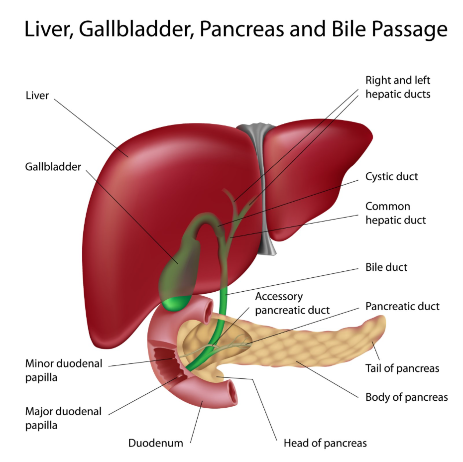
In cases of intra-hepatic jaundice, such as hepatitis, the bile ducts will usually appear normal on a scan, while the liver tissue itself might look slightly swollen or “bright.” In post-hepatic cases, the ducts will be dilated because bile is backing up behind a stone or tumour. By using these visual clues, the healthcare team can quickly decide if the patient needs to see a hepatologist for liver disease or a surgeon for a biliary obstruction.
Identifying Gallstones and Duct Obstructions
A primary use of imaging in jaundice is the identification of gallstones that have migrated into the common bile duct. While gallstones in the gallbladder are common and do not always cause jaundice, a stone lodged in the duct prevents bilirubin from reaching the intestines. NICE clinical guidelines for gallstone disease management state that imaging should be used to confirm the presence of stones in the bile duct when symptoms like jaundice and pain are present.
Both ultrasound and CT scans have specific roles here. Ultrasound is the gold standard for looking at the gallbladder, but if a stone is very deep in the duct, it can be harder to see. In such cases, a CT scan or a more specialised MRI scan might be used to pinpoint the exact location of the obstruction. Identifying the “level” of the blockage—whether it is high up near the liver or low down near the pancreas—is essential for planning the correct procedure to remove it and restore normal bile flow.
Preparation and Process for Jaundice Scans
In the United Kingdom, patients are given specific instructions to prepare for an ultrasound or CT scan to ensure the images are as clear as possible. For an abdominal ultrasound, patients are usually asked to fast (not eat or drink anything except water) for at least six hours before the appointment. This allows the gallbladder to remain full of bile, making it easier to see any stones inside.
For a CT scan, preparation might involve drinking a specific fluid (oral contrast) or having an intravenous injection of contrast dye during the procedure. Patients must inform the clinical team if they have any allergies or kidney problems before receiving contrast. Both procedures are relatively quick; an ultrasound usually takes 15 to 20 minutes, while a CT scan may only take a few minutes once the patient is positioned on the scanner table. These structured processes ensure that the diagnostic pathway is efficient and that the multidisciplinary team receives the high-quality images they need to make a definitive assessment.
Specialist Imaging: Beyond Ultrasound and CT
If ultrasound and CT scans are inconclusive but the clinical suspicion of a blockage remains high, UK specialists may request a specialised MRI known as an MRCP (Magnetic Resonance Cholangio-Pancreatography). This scan uses powerful magnets and radio waves to create a detailed three-dimensional map of the bile ducts and pancreatic duct. It is a highly sensitive, non-invasive way to find small stones or narrowing that other scans might miss.
The GOV.UK health pages indicate that advanced imaging is a vital part of the UK’s strategy for the early diagnosis of biliary and liver malignancies. In some cases, a procedure called an ERCP may follow a scan, where a camera is passed down the throat to physically remove a blockage. By following this sequential imaging pathway—starting with ultrasound and moving to CT or MRI if necessary—the NHS ensures that patients receive the most appropriate investigation for their specific symptoms, leading to an accurate and timely diagnosis.
Conclusion
Ultrasound and CT scans are essential tools for diagnosing the cause of jaundice by providing a visual assessment of the liver, gallbladder, and bile ducts. Ultrasound is typically the first step for identifying gallstones and duct dilation, while CT scans offer more detail for assessing tumours or complex liver changes. In the UK, these imaging results guide clinicians in determining whether the jaundice is caused by an internal liver issue or a physical obstruction requiring intervention. Understanding the purpose of these scans allows for a clearer diagnostic journey and helps ensure that the correct management is initiated. If you experience severe, sudden, or worsening symptoms, call 999 immediately.
Does a normal ultrasound mean I don’t have liver disease?
Not necessarily; some liver conditions like hepatitis do not change the liver’s physical appearance enough to show up on an ultrasound, so blood tests remain vital.
Why can’t I just have a CT scan instead of an ultrasound?
Ultrasound is actually better and safer for finding gallstones, while CT is better for looking at larger structures and finding tumours.
Will the contrast dye for a CT scan make me feel sick?
Some people feel a brief sensation of warmth or a metallic taste in the mouth, but significant reactions are rare and monitored by the staff.
Can a scan tell if my jaundice is from alcohol?
A scan can show if your liver is fatty or scarred, which might suggest alcohol-related damage, but a medical history is needed for confirmation.
How soon after the scan will I get the results?
In most UK hospitals, the images are reviewed by a radiologist and the report is sent back to your doctor within a few days, or sooner if urgent.
Is there any radiation in an ultrasound?
No, ultrasound uses sound waves only, making it a very safe option with no radiation risk.
What is the difference between an MRCP and a standard MRI?
An MRCP is a specific type of MRI scan that focuses entirely on the bile ducts and gallbladder to look for blockages or stones.
Authority Snapshot (E-E-A-T)
This article provides medically factual health education regarding the use of imaging in jaundice diagnosis, strictly aligned with NHS and NICE clinical guidelines. The content is developed by a professional medical writing team and reviewed by Dr. Stefan Petrov, a UK-trained physician with experience in general medicine, surgery, and emergency care. All information follows current UK public health protocols to ensure accuracy and patient safety.