




Inflammation from diverticulitis can significantly obscure the presence of colon polyps on various types of medical imaging by causing the bowel wall to thicken and creating physical distortions that hide small tissue growths. When the small pouches in the colon wall, known as diverticula, become infected or inflamed, the surrounding area undergoes a rapid immune response involving swelling, increased blood flow, and the accumulation of fluid. This localised reaction transforms the usually smooth and thin lining of the large intestine into a thickened, irregular surface that can mask the subtle protrusions of a polyp. In the United Kingdom, healthcare professionals recognise that an acute flare-up of diverticulitis creates a suboptimal environment for a thorough visual or radiological inspection of the bowel. Consequently, standard clinical protocols often involve treating the infection first and waiting for the inflammation to subside before performing definitive screenings for other structural abnormalities. Understanding the biological and technical reasons why inflammation interferes with imaging is essential for patients navigating the diagnostic process. By ensuring that the colon is inspected only after the tissue has returned to its healthy state, the UK healthcare system maintains high standards of accuracy in detecting and preventing serious bowel conditions.
What We’ll Discuss in This Article
- The biological impact of bowel wall thickening on diagnostic clarity.
- How swelling and fluid accumulation physically mask colon polyps.
- Limitations of CT scans and ultrasounds during active inflammatory episodes.
- The clinical necessity of delaying visual inspections like colonoscopies.
- Structural differences between inflammatory swelling and tissue overgrowths.
- UK clinical pathways for ensuring a clear colon after an infection.
Biological Wall Thickening and Diagnostic Obscurity
Active inflammation from diverticulitis causes the colon wall to become significantly thicker, which can bury small polyps within the swollen folds of the intestinal lining. In a healthy state, the bowel wall is relatively thin and flexible, allowing imaging tools to clearly define the boundary between the tissue and the internal space where waste passes. However, during an acute flare-up, the body’s inflammatory response leads to oedema, which is an accumulation of fluid within the tissue layers. The NHS states that diverticulitis happens when the small pouches in the lining of the large intestine become inflamed or infected, often causing the bowel wall to swell.
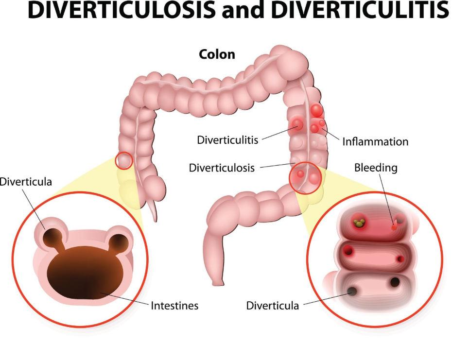
This swelling causes the mucosal lining to bulge and create extra folds, known as haustral thickening. A small or flat polyp can easily be lost within these deep, inflamed ridges, making it invisible to a radiologist or endoscopist. Because the goal of imaging is to find growths that might require removal, performing these checks while the wall is in a reactive state significantly increases the risk of a “false negative” result, where a polyp is present but cannot be seen. In the UK, clinicians wait for this thickening to resolve to ensure that every millimetre of the lining can be accurately inspected.
Physical Masking by Fluid and Mucus
The presence of inflammatory fluids, mucus, and localised debris during a diverticulitis flare-up provides a physical barrier that prevents imaging tools from getting a clear view of the underlying colon polyps. Inflammation triggers the goblet cells in the bowel lining to produce excess mucus as a protective measure, which can coat the surface of the intestine.
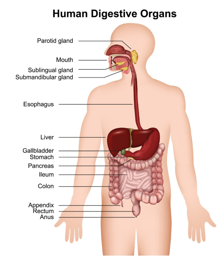
On a radiological scan or during a visual examination, this thick layer of fluid and mucus can smooth over the appearance of a small polyp or be mistaken for the wall itself. Furthermore, diverticulitis can lead to “fat stranding,” where the fat surrounding the colon becomes inflamed and cloudy on a scan. This cloudiness reduces the contrast between the bowel wall and the surrounding structures, making it even harder for doctors to identify small protrusions like polyps. In the United Kingdom, resolving this “noisy” environment is a prerequisite for a reliable bowel check. By allowing the infection to clear, the mucosal surface becomes clean and dry again, providing the high-definition clarity required to spot subtle tissue changes.
Limitations of Radiological Scans During Inflammation
Computed Tomography (CT) scans, while excellent for diagnosing diverticulitis, are limited in their ability to detect polyps when the colon is actively inflamed because the swelling mimics the appearance of other structural issues. A CT scan uses X-rays to create cross-sectional images, but the density of an inflamed bowel wall is very similar to the density of a fleshy polyp.
| Feature | Diverticulitis Flare-up on Scan | Healthy Colon on Scan |
| Wall Appearance | Thickened, blurred, and irregular. | Thin, crisp, and well-defined. |
| Visual Contrast | Low; obscured by fluid and fat stranding. | High; clear boundary between tissue and air. |
| Polyp Visibility | Poor; can be hidden by wall swelling. | High; growths clearly protrude into lumen. |
| Procedural Risk | Low risk (non-invasive). | Low risk (non-invasive). |
Because of these technical limitations, a CT scan performed during a flare-up is primarily used to check for complications like abscesses rather than to screen for polyps. NICE clinical guidelines for diverticular disease management identify CT as the gold standard for diagnosing acute diverticulitis but caution that it may not be suitable for ruling out other structural pathologies during the acute phase. In UK clinical practice, if a scan shows significant inflammation, it is standard to recommend a follow-up visual check later to ensure no polyps were missed while the wall was swollen.
Safety and Clarity: The Case for Delaying Colonoscopy
Invasive visual inspections like a colonoscopy are delayed after diverticulitis because the inflammation makes the bowel wall too fragile for a safe procedure and too obscured for an accurate one. A colonoscopy requires the bowel to be inflated with air or carbon dioxide to smooth out the folds and provide a clear view. During an active infection, the pressure from this inflation could cause a weakened diverticulum to perforate or burst.
Beyond the safety risk, the visual environment during diverticulitis is often filled with redness, swelling, and pus, which makes it impossible for the endoscopist to be certain they have seen everything. The GOV.UK health pages indicate that follow-up investigations to rule out polyps or other conditions are typically scheduled six to eight weeks after a diverticulitis flare-up has resolved. This recovery period allows the “reactive” tissue to return to normal, ensuring that the bowel wall is strong enough for the procedure and clean enough for a definitive inspection. By following this staged approach, UK healthcare providers ensure that polyps are not missed due to the temporary distortions caused by infection.
Differentiating Inflammatory Swelling from Tissue Overgrowth
Clinicians distinguish between inflammatory swelling and colon polyps by looking for specific patterns of tissue change, a task that becomes much easier once the acute infection has passed. Inflammatory thickening is usually “circumferential,” meaning it affects the entire ring of the bowel wall in a certain section. In contrast, polyps are typically “focal,” appearing as a single, isolated bump or stalk on an otherwise healthy area of the lining.
During a follow-up scan or colonoscopy, the absence of widespread redness and swelling allows these focal growths to stand out clearly. If a protrusion is found after the diverticulitis has cleared, the clinician can be much more confident that it is a true polyp and not just a swollen fold of tissue. This distinction is vital for determining the next steps in care, such as a polypectomy (removal of the growth). In the UK, this process of elimination is a cornerstone of gastrointestinal diagnostic safety, preventing unnecessary surgeries for swelling while ensuring that high-risk polyps are identified and removed.
UK Clinical Protocols for Post-Infection Monitoring
The United Kingdom uses integrated clinical pathways to ensure that every patient who has experienced diverticulitis eventually receives a clear and accurate check for polyps. Once the acute symptoms like pain and fever have subsided, the patient is usually scheduled for a review to plan their follow-up investigation.
This protocol ensures that:
- The bowel wall is healed: Reducing the risk of perforation during an invasive check.
- Inflammation markers are normal: Checking blood tests to ensure the body is no longer in a reactive state.
- Diagnostic accuracy is maximised: Providing the best possible environment to find and remove polyps.
- Long-term risk is managed: Transitioning the patient from acute infection care back to preventative screening.
By following this evidence-based sequence, the NHS provides a comprehensive safety net. It addresses the immediate crisis of diverticulitis while maintaining a vigilant focus on the long-term goal of identifying cellular overgrowths. Patients are encouraged to attend these follow-up appointments even if they feel completely better, as it is the only way to be certain that the inflammation was not hiding a “silent” but potentially significant growth in the large intestine.
Conclusion
Inflammation from diverticulitis can hide colon polyps on imaging by thickening the bowel wall and creating physical distortions through swelling and mucus. While CT scans are effective for diagnosing the infection, they are less reliable for spotting small growths during the acute phase. In the UK, clinicians wait for the tissue to heal before performing definitive follow-up checks like a colonoscopy to ensure safety and diagnostic accuracy. This staged approach ensures that both the infectious and cellular risks to the colon are managed effectively. If you experience severe, sudden, or worsening symptoms, call 999 immediately.
Can a CT scan tell the difference between a polyp and swelling?
During a flare-up, it can be very difficult to tell them apart because they have similar densities; this is why a follow-up check is needed once the swelling goes down.
What happens if a scan misses a polyp because of inflammation?
This is why UK doctors recommend a follow-up colonoscopy six to eight weeks later; it serves as a “double-check” to find anything that was hidden.
Is a colonoscopy more accurate than a CT scan for finding polyps?
Yes, because it allows the clinician to see the lining directly in high definition and move folds of tissue to see what is behind them.
Will my diverticulitis pain make it harder to find polyps?
Indirectly, yes; if you are in pain, you cannot have the invasive tests needed to find polyps safely, so the infection must be treated first.
Does everyone who has diverticulitis need a follow-up check for polyps?
In the UK, it is standard practice for most people after their first flare-up to ensure the diagnosis was correct and the colon is otherwise healthy.
Can a home screening kit find polyps if I have inflammation?
No; the home kit only looks for hidden blood and cannot see through swelling or inflammation to identify structural issues.
How do I know when my inflammation has gone down enough for a scan?
Your doctor will check your symptoms and may use a blood test to see if your inflammatory markers (like CRP) have returned to a normal level.
Authority Snapshot (E-E-A-T)
This article provides medically factual health education regarding the impact of diverticulitis on bowel imaging, strictly aligned with NHS and NICE clinical guidelines. The content is developed by a professional medical writing team and reviewed by Dr. Stefan Petrov, a UK-trained physician with extensive experience in emergency care, surgery, and general medicine. All information follows current UK public health protocols to ensure clinical accuracy and patient safety.



