
When a patient presents with symptoms such as breathlessness or persistent fatigue, doctors follow a structured diagnostic pathway to determine if the heart is struggling to pump blood efficiently. This process involves a combination of clinical history, physical examinations, and specific diagnostic tests designed to evaluate the heart’s structure and function. Identifying the specific type of heart failure is essential because it informs the medical team about how the heart muscle is behaving and which evidence-based treatments will be most effective for the individual’s condition.
What We’ll Discuss in This Article
- The initial clinical assessments and blood tests used in the UK.
- The role of the echocardiogram in categorising heart failure.
- Understanding the concept of ejection fraction.
- The difference between heart failure with reduced and preserved function.
- How diagnostic results guide the specialist referral process.
- Additional investigations that may be required for specific cases.
The initial diagnostic steps and blood testing
The diagnostic journey typically begins in a primary care setting where a GP assesses your symptoms and medical history. Doctors look for signs of fluid retention, such as swollen ankles or lungs that sound congested during a physical examination. If heart failure is suspected, the first formal investigation is almost always a specific blood test called an NT-proBNP test. This test measures a protein that the heart releases when it is under increased pressure or stretch.
According to NHS guidance on heart failure diagnosis, the results of this blood test determine the urgency of further investigations. If the levels of NT-proBNP are high, it indicates that the heart may be struggling, prompting a referral to a specialist for more detailed imaging. This blood test is a vital screening tool because a normal result usually means that heart failure is very unlikely, allowing doctors to explore other potential causes for your symptoms without delay.
The role of the echocardiogram
The primary tool used to categorise the type of heart failure is a transthoracic echocardiogram, which is an ultrasound scan of the heart. This non-invasive procedure allows a cardiologist or a specialist sonographer to see the heart’s chambers in motion, check the health of the heart valves, and measure how much blood is being pumped out with each contraction. The information gathered during this scan is the most important factor in determining the specific subtype of heart failure.
During the echocardiogram, the specialist focuses on the left ventricle, which is the heart’s main pumping chamber. By observing the thickness of the heart walls and the size of the chambers, the medical team can identify if the heart muscle has become weakened and stretched or if it has become stiff and thickened. These structural changes are the physical markers that doctors use to differentiate between the various forms of the condition.
Understanding Ejection Fraction (EF)
Ejection fraction is a measurement expressed as a percentage that describes how much of the blood in the left ventricle is pumped out with each heartbeat. In a healthy heart, the ejection fraction is typically between 50 per cent and 70 per cent. Doctors use this percentage as a primary metric to classify heart failure into distinct categories. This classification is not just a label. It is a clinical tool that determines which medications, such as ACE inhibitors or beta-blockers, are likely to provide the most benefit.
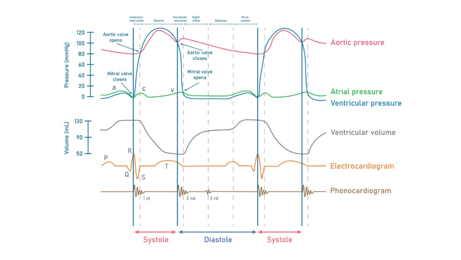
The NICE guidance for chronic heart failure provides the framework for using these measurements. If the ejection fraction is low, usually below 40 per cent, the condition is classified in a way that suggests the heart muscle is not contracting with enough force. If the percentage is higher but the patient still has symptoms, it indicates a different mechanical issue, usually related to how the heart relaxes and fills with blood.
Categorising the specific types of heart failure
Once the echocardiogram and ejection fraction data are available, doctors can place the condition into one of several categories. The two most common types are Heart Failure with Reduced Ejection Fraction (HFrEF) and Heart Failure with Preserved Ejection Fraction (HFpEF). While the symptoms for both can be identical, the underlying physiology of the heart is different in each case.
| Heart Failure Type | Pumping Function | Description |
| Reduced (HFrEF) | Weakened contraction | The heart muscle is thin or weak and cannot squeeze enough blood out. |
| Preserved (HFpEF) | Impaired filling | The heart muscle is stiff or thick and cannot relax enough to fill with blood. |
| Mildly Reduced (HFmrEF) | Borderline | The pumping function falls into a middle range between reduced and preserved. |
In HFrEF, the heart has become enlarged and the muscle is less able to push blood into the circulation. In HFpEF, the heart can still pump relatively well, but because the muscle is stiff, it cannot hold enough blood to meet the body’s needs. Distinguishing between these is the core objective of the diagnostic process.
Further investigations for complex cases
In some instances, the initial blood tests and ultrasound may not provide a complete picture, and further investigations are required to understand the cause of the heart failure. This might include a cardiac MRI, which provides even more detailed images of the heart tissue, or a coronary angiogram to check if the heart’s blood supply is restricted. These tests help doctors determine if the heart failure was caused by a previous heart attack, high blood pressure, or a problem with the heart valves.
Doctors may also perform an electrocardiogram (ECG) to check the heart’s electrical rhythm. Rhythm disturbances, such as atrial fibrillation, can contribute to heart failure or be a result of it. By identifying these additional factors, the specialist team can create a comprehensive management plan that addresses both the pumping function of the heart and any underlying issues that are making the condition worse.
Conclusion
Doctors work out the type of heart failure by following a rigorous, evidence-based pathway that starts with symptoms and blood tests and leads to detailed heart imaging. The echocardiogram remains the most vital tool in this process, providing the ejection fraction measurement that allows for clear clinical categorisation. Understanding whether the heart is struggling to contract or struggling to fill is the essential first step in ensuring patients receive the specific treatments recommended by UK national guidelines.
If you experience severe, sudden, or worsening symptoms, call 999 immediately.
FAQ Section
Is one type of heart failure more serious than the other?
Both types require careful medical management and can significantly impact a person’s daily life, but they are treated with different sets of medications. Your specialist will explain the outlook based on your specific results and overall health.
Can the type of heart failure I have change over time?
Yes, with effective treatment and lifestyle changes, some patients may see an improvement in their ejection fraction, which might change how their condition is categorised.
Why did I need a blood test before I could get a heart scan?
The blood test acts as a screening tool to ensure that those with the highest probability of heart failure are prioritised for specialist scans as quickly as possible.
Will I need to have an echocardiogram more than once?
Specialists often repeat the scan at certain intervals to see how the heart is responding to medication or to monitor any changes in the heart’s structure.
Does my age affect how these tests are interpreted?
Doctors take age into account because certain heart measurements and blood test results can change naturally as people get older.
What is the difference between an ECG and an echocardiogram?
An ECG records the electrical activity and rhythm of the heart, while an echocardiogram uses ultrasound to look at the physical structure and pumping action of the heart.
Can I have heart failure if my heart looks normal on a scan?
It is possible to have symptoms and heart strain even if the heart’s pumping action appears normal, which is why doctors look at the heart’s ability to relax as well as its ability to squeeze.
Authority Snapshot
This article is designed to assist patients in understanding the UK diagnostic process for heart failure. It was written by Dr. Rebecca Fernandez, a UK-trained physician with clinical experience in cardiology and internal medicine. All information provided is strictly aligned with current NHS and NICE clinical guidelines to ensure patient safety and accuracy.
