A metastatic brain tumour, also commonly referred to as a secondary brain tumour, is a growth that develops when cancer cells spread to the brain from another part of the body. Unlike primary brain tumours that originate within the neurological tissue itself, metastatic tumours are composed of cells from the original primary cancer site, such as the lungs or breasts. In the United Kingdom, healthcare professionals manage these conditions through integrated oncology pathways that address both the primary disease and its spread to the central nervous system. This article provides a clinical overview of how metastatic tumours form and the standardised processes used by the NHS and NICE to coordinate specialist care. Understanding the biological nature of metastasis is essential for patients and families as they navigate the diagnostic and management journey within the UK healthcare system.
What We’ll Discuss in This Article
- The biological process of cancer spreading to the brain from primary sites.
- Common primary cancers that are most likely to result in brain metastases.
- The difference between primary and secondary brain tumours.
- Symptoms associated with metastatic growths and increased intracranial pressure.
- The diagnostic process in the UK using MRI imaging and full body staging.
- Integrated management pathways involving surgery, radiation, and systemic therapy.
The Biological Process of Brain Metastasis
Metastatic brain tumours develop when cancer cells break away from a primary tumour in another organ and travel through the bloodstream or lymphatic system to reach the brain. Once these cells arrive in the small blood vessels of the brain, they can migrate into the brain tissue and begin to multiply, forming one or more new tumours. The NHS states that a metastatic brain tumour is cancer that has spread to the brain from another part of the body.
This process is complex because the cells must bypass the blood-brain barrier, a protective shield that normally prevents harmful substances from entering the neurological environment. In the United Kingdom, clinicians use the term “secondary” because the cancer is not new to the brain; if lung cancer spreads to the brain, the cells in the brain are still lung cancer cells. This biological distinction is vital for determining the most effective management, as the cells often remain sensitive to the same systemic therapies used for the original primary site.
Common Primary Sources of Metastasis
While any cancer can potentially spread to the brain, certain types are statistically more likely to develop metastases, including lung, breast, kidney, and skin cancers. In the United Kingdom, lung cancer is the most common source of secondary brain tumours, followed closely by breast cancer and melanoma.
| Primary Cancer Site | Likelihood of Brain Spread | Common Characteristics |
| Lung Cancer | High | Often present at the time of initial diagnosis. |
| Breast Cancer | Moderate | May occur several years after the primary diagnosis. |
| Melanoma | High | Can spread quickly and often involves multiple sites. |
| Kidney Cancer | Moderate | Frequently results in highly vascular tumours. |
| Bowel Cancer | Low | Less common but can occur in advanced stages. |
The frequency of brain metastasis has increased in recent years because UK patients are living longer with their primary cancers due to improved systemic treatments. This extended survival period provides a longer window of time for cells to eventually reach the brain. Understanding the primary source helps UK multidisciplinary teams predict the behaviour of the secondary growths and choose the most appropriate targeted therapies.
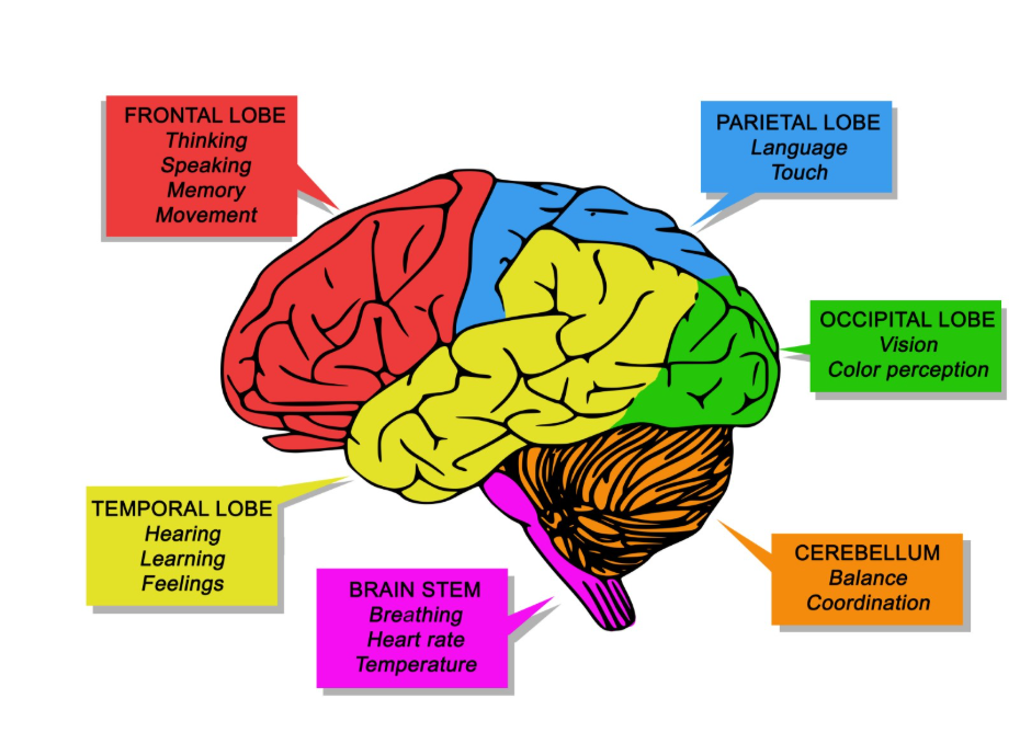
Distinguishing Primary and Secondary Tumours
In the UK clinical setting, it is essential to distinguish between primary and secondary brain tumours because their biological drivers and management strategies are entirely different. A primary tumour starts in the brain cells, supportive glial cells, or the membranes surrounding the brain, whereas a secondary tumour is an invasive visitor from another organ.
NICE clinical guidelines for brain tumours indicate that the management of metastatic disease must be coordinated between the primary cancer site specialists and the neuro-oncology team. Primary tumours are often single masses, while metastatic disease frequently involves multiple tumours in different parts of the brain. Additionally, metastatic tumours often cause significant “oedema” or swelling in the surrounding healthy brain tissue. UK radiologists look for specific imaging features, such as the location and the way the tumour absorbs contrast dye, to help differentiate between the two types during the initial assessment.
Common Symptoms and Mass Effect
Symptoms of a metastatic brain tumour are caused by the physical presence of the mass and the associated swelling, which increase the pressure within the rigid structure of the skull. This is often referred to as “mass effect” and can lead to both general symptoms of pressure and focal symptoms related to the specific part of the brain affected.
The GOV.UK health pages provide clinical profiles indicating that common symptoms of a secondary brain tumour include persistent headaches, new-onset seizures, and changes in personality or vision. Headaches associated with metastasis are often worse in the morning or when bending over and may be accompanied by nausea. Focal symptoms might include weakness on one side of the body, difficulty speaking, or a loss of coordination. Because metastatic tumours can occur in multiple locations, a patient might experience several different neurological symptoms simultaneously. In the UK, if a patient with a known history of cancer develops new neurological signs, they are prioritised for urgent brain imaging.
The Diagnostic Process in the UK
The diagnostic pathway for a suspected metastatic brain tumour in the United Kingdom involves high-resolution MRI imaging of the brain and “staging” scans of the rest of the body to identify the primary source. If a patient is not already known to have cancer, finding a brain tumour often triggers a search for a primary tumour elsewhere, such as a CT scan of the chest, abdomen, and pelvis.
An MRI with contrast dye is the preferred tool for identifying metastases because it is highly sensitive at detecting even very small tumours that might be missed on a CT scan. The diagnostic process is coordinated through a regional neuro-oncology multidisciplinary team (MDT), which includes neurosurgeons, oncologists, and radiologists. If the primary source is unknown, a biopsy of the brain tumour or a more accessible site elsewhere may be required to confirm the cell type. This integrated approach ensures that the clinical team has a complete picture of the disease throughout the body before starting a management plan.
Integrated Management Pathways
Management of metastatic brain tumours in the United Kingdom is highly personalised and depends on the number of tumours, the status of the primary cancer, and the patient’s overall health. The goal of management is often to control the neurological symptoms and preserve quality of life while continuing to address the systemic disease.

The UK management approach includes:
- Steroids: Using medication like dexamethasone to rapidly reduce the swelling around the tumours.
- Surgery: Physically removing a single, large tumour if it is causing significant symptoms or pressure.
- Radiotherapy: Utilizing targeted beams or whole-brain treatment to destroy abnormal cells.
- Stereotactic Radiosurgery (SRS): Delivering precise, high-dose radiation to small, specific tumours.
- Systemic Therapy: Using targeted drugs or immunotherapy that can cross the blood-brain barrier.
In the UK, many patients are now managed with SRS, which allows for precise treatment of multiple small tumours while sparing healthy brain tissue. This technology has significantly improved the ability of the NHS to manage secondary brain disease. Throughout the process, patients receive support from specialist nurses and palliative care teams who focus on symptom control and holistic wellbeing.
Conclusion
A metastatic brain tumour is a secondary growth that has spread from a primary cancer elsewhere in the body and requires a coordinated clinical response. In the UK, the NHS uses advanced MRI imaging and multidisciplinary reviews to manage these tumours while prioritising the patient’s neurological function and quality of life. Management options include surgery, targeted radiation, and systemic therapies tailored to the original primary cancer type. Consistent monitoring and expert support are essential for navigating the complexities of metastatic disease. If you experience severe, sudden, or worsening symptoms, call 999 immediately.
Is a metastatic brain tumour the same as brain cancer?
It is cancer in the brain, but it is classified and managed as the primary cancer; for example, it is called metastatic lung cancer, not primary brain cancer.
Can a metastatic brain tumour be cured?
While management focuses on control and symptom relief, modern treatments like radiosurgery can effectively manage individual tumours for long periods.
Why do I have multiple tumours in the brain?
Metastatic cells often travel in groups through the bloodstream, which frequently leads to the development of more than one tumour in different areas.
Does having a brain tumour mean the cancer is everywhere?
Not necessarily; metastasis to the brain is a specific stage of spread that requires targeted management alongside the primary site.
Will radiotherapy make my hair fall out?
Whole-brain radiotherapy can cause hair loss, but modern targeted stereotactic radiosurgery usually only affects hair in a very small, specific area.
Can medication shrink the tumours without surgery?
Yes, some modern targeted therapies and immunotherapies are very effective at crossing into the brain and shrinking metastatic tumours.
What is the difference between a CT and an MRI for these tumours?
MRI provides much clearer detail of the brain tissue and is better at finding small metastatic tumours that a CT scan might miss.
Authority Snapshot (E-E-A-T)
This article provides medically factual health education regarding metastatic brain tumours, strictly aligned with NHS and NICE clinical guidelines. The content is developed by a professional medical writing team and reviewed by Dr. Stefan Petrov, a UK-trained physician with experience in surgery, emergency care, and clinical education. All information follows current UK public health protocols to ensure clinical accuracy and patient safety.
