The detection of a brain tumour in the United Kingdom primarily relies on advanced neuroimaging techniques, specifically Magnetic Resonance Imaging and Computed Tomography scans, to visualise the internal structures of the skull. These scans allow healthcare professionals to identify abnormal growths, assess their size and location, and determine their relationship with surrounding healthy brain tissue. In the UK, the choice of scan is determined by the clinical urgency and the specific information required by the neurosurgical team. By adhering to evidence-based protocols established by the NHS and NICE, clinical teams ensure that patients receive the most appropriate imaging for an accurate and timely diagnosis. Understanding the different types of scans and their specific roles is essential for patients as they move through the diagnostic pathway within the UK healthcare system.
What We’ll Discuss in This Article
- The role of Magnetic Resonance Imaging as the primary diagnostic tool.
- How Computed Tomography is utilised in emergency clinical settings.
- The importance of contrast agents in highlighting tumour characteristics.
- Specialized imaging techniques such as PET scans and MRS.
- The UK clinical pathway for referral and access to neuroimaging.
- Safety considerations and what to expect during a brain scan.
Magnetic Resonance Imaging (MRI) as the Gold Standard
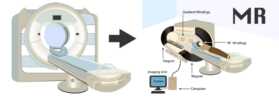
Magnetic Resonance Imaging is the primary scan used to detect and evaluate brain tumours in the United Kingdom because it provides superior detail of soft tissues without using ionising radiation. An MRI uses strong magnetic fields and radio waves to produce high-resolution, multi-dimensional images of the brain. The NHS states that an MRI scan is the main type of scan used to diagnose a brain tumour as it provides a very detailed picture of the inside of the brain.
This technology is particularly effective at identifying gliomas, meningiomas, and other primary tumours that may have subtle borders. UK clinicians prefer MRI because it can distinguish between different types of tissue, such as swelling (oedema), tumour mass, and healthy grey or white matter. Because the scan takes longer than a CT, it requires the patient to lie still for approximately 30 to 60 minutes. The level of detail provided by an MRI is essential for the multidisciplinary team to plan surgical interventions or targeted radiation treatments. In the UK, the NHS prioritises MRI for any non-emergency neurological investigation where a structural issue is suspected.
Computed Tomography (CT) in Emergency Assessment
Computed Tomography scans are frequently used as the initial imaging tool in emergency departments across the United Kingdom when a patient presents with sudden or severe neurological symptoms. A CT scan uses a series of X-rays and computer processing to create cross-sectional images of the head. NICE clinical guidelines for brain tumours indicate that a CT scan may be performed first in an emergency to quickly rule out other causes of symptoms, such as a brain haemorrhage or a large mass causing significant pressure.
| Feature | MRI Scan | CT Scan |
| Technology | Magnetic fields and radio waves. | X-rays and computer processing. |
| Duration | 30 to 60 minutes. | 5 to 10 minutes. |
| Detail Level | High detail of soft brain tissue. | Best for bone and acute bleeding. |
| Radiation | No ionising radiation used. | Uses ionising radiation. |
| Suitability | Definitive tumour diagnosis. | Emergency triage and screening. |
While a CT scan is faster and more accessible than an MRI, it provides less detail regarding the internal cellular structure of a tumour. However, it is highly effective at showing changes in the skull bone or the presence of calcium within a tumour. In the UK, if a CT scan identifies a suspicious area, it is almost always followed by a more detailed MRI to provide the neurosurgeon with the information needed for a definitive management plan.
The Role of Contrast Agents in Neuroimaging
Contrast agents, often referred to as dyes, are frequently used in both MRI and CT scans in the United Kingdom to make a brain tumour stand out more clearly against healthy tissue. These agents, typically containing gadolinium for MRI or iodine for CT, are injected into a vein in the arm shortly before or during the scan. The GOV.UK health pages provide clinical profiles indicating that contrast-enhanced imaging is a standard requirement for assessing the vascularity and malignancy of a suspected brain tumour.
Malignant or high-grade tumours often have a disrupted blood-brain barrier and a high number of blood vessels, causing them to absorb more contrast and appear “bright” on the final images. This “enhancement” helps radiologists determine the most active parts of a tumour, which is vital for choosing the best site for a biopsy. In the UK, medical teams screen patients for kidney function or allergies before administering contrast to ensure the procedure is safe. By using contrast, UK specialists can more accurately grade a tumour and monitor its response to treatment over time.
Specialized Scans: PET and MRS
In some complex cases, UK specialists may utilise specialised scans like Positron Emission Tomography or Magnetic Resonance Spectroscopy to gain deeper insights into the tumour’s biological activity. A PET scan involves injecting a small amount of a radioactive tracer that highlights areas of high metabolic activity, which can help distinguish between active tumour cells and scar tissue from previous treatments.
Magnetic Resonance Spectroscopy (MRS) is a non-invasive technique that can be performed during a standard MRI session. It measures the chemical metabolites within a specific area of the brain, providing a “biochemical fingerprint” of the tissue. This can help clinicians determine the likely grade of a tumour without an immediate biopsy. While these specialised scans are not part of the initial screening for everyone in the UK, they are essential tools for multidisciplinary teams when standard imaging results are inconclusive. These technologies represent the advanced diagnostic capabilities available within the NHS to ensure precise neurological care.
Safety and What to Expect During Scans
Safety is a primary concern in the UK healthcare system, and strict protocols are followed to ensure that every patient is prepared and protected during their neuroimaging session. For an MRI, patients must undergo a thorough screening to ensure they do not have any metal implants, such as pacemakers or certain types of surgical clips, that could be affected by the strong magnetic field.
Patients can expect the following during their scan:
- Preparation: Removing jewellery, watches, and clothing with metal fasteners.
- Positioning: Lying flat on a motorised table that moves into the scanner.
- Noise: MRI scanners are very noisy, so patients are provided with earplugs or headphones.
- Communication: Being able to speak to the radiographer via an intercom throughout the procedure.
- Contrast Injection: A small needle may be placed in the arm if dye is required.
In the UK, radiographers are highly trained to support patients who may feel claustrophobic or anxious during the process. If a patient is unable to tolerate the enclosed space of a standard MRI, some NHS trusts offer access to “open” or wider-bore scanners. Ensuring patient comfort and safety allows for the collection of high-quality images, which is the foundation of an accurate neurological diagnosis.
UK Clinical Pathways for Imaging Referrals
The United Kingdom has established integrated care pathways to ensure that patients with suspected brain tumours receive their scans within a specific timeframe. Most patients are referred by their GP under the “two-week wait” or the 28-day faster diagnosis standard if they present with “red flag” symptoms such as new seizures or persistent morning headaches.
The UK diagnostic imaging pathway involves:
- Clinical Triage: Ensuring the most appropriate scan (MRI or CT) is requested based on symptoms.
- Reporting: Specialist neuroradiologists interpreting the images to identify structural abnormalities.
- Communication: Results being sent to the referring consultant or the multidisciplinary team.
- Follow-up: Scheduling additional scans if the initial results require further investigation.
This structured system ensures that serious conditions are identified as quickly as possible, which is vital for preserving neurological function. By following these national standards, the NHS provides a consistent level of diagnostic care across all regions. Once a scan has detected a tumour, the images are used as a roadmap for the subsequent biopsy or surgical intervention, ensuring that the clinical journey is guided by the most accurate data available.
Conclusion
Detecting a brain tumour involves a structured pathway of imaging, with MRI serving as the gold standard for detailed diagnosis and CT utilised for rapid emergency assessments. In the UK, the NHS follows strict protocols using contrast agents and specialised techniques like PET scans to provide a comprehensive biological profile of any detected mass. These scans are essential for planning surgery and monitoring the effectiveness of management over time. Understanding the role of each scan helps patients participate actively in their diagnostic journey while ensuring their safety is prioritised. If you experience severe, sudden, or worsening symptoms, call 999 immediately.
Can a brain tumour be missed on a CT scan?
Yes; some small or low-grade tumours may not show up clearly on a CT, which is why a follow-up MRI is often required if symptoms persist.
Is the dye used in scans dangerous?
Contrast agents are generally safe, but UK clinicians check your kidney function and allergy history before use to minimise any risk.
Why can’t I have an MRI if I have a pacemaker?
The strong magnets in the MRI can interfere with the electronic components of older pacemakers, although some newer models are MRI-compatible.
Will a brain scan show why I have headaches?
A scan can show if a structural issue like a tumour is causing headaches, but most headaches are not related to tumours and may have other causes.
How long do I have to wait for scan results in the UK?
While the scan itself is fast, it usually takes a few days for a specialist radiologist to write the report and for your doctor to review it.
Are there any side effects from a brain scan?
A CT scan involves a small dose of radiation, while an MRI has no known side effects, though some people feel slightly claustrophobic.
Can children have these scans?
Yes; children can have both CT and MRI scans, though they may sometimes require a sedative to help them stay still for the duration of the procedure.
Authority Snapshot (E-E-A-T)
This article provides medically factual health education regarding brain tumour scans, strictly aligned with NHS and NICE clinical guidelines. The content is developed by a professional medical writing team and reviewed by Dr. Stefan Petrov, a UK-trained physician with experience in surgery, emergency care, and clinical education. All information follows current UK public health protocols to ensure clinical accuracy and patient safety.
